
Dermis Collection
The dermis, a fascinating layer of our skin, plays a crucial role in protecting and maintaining our body


All Professionally Made to Order for Quick Shipping
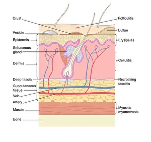
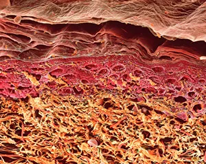
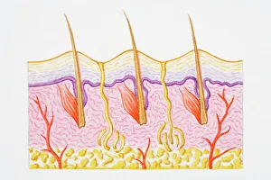
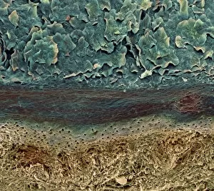
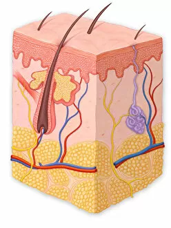
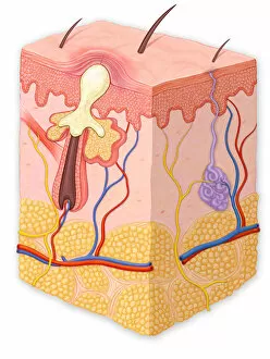
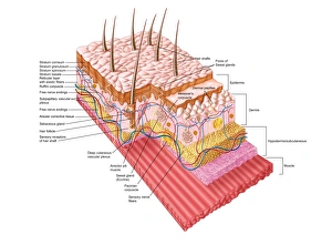
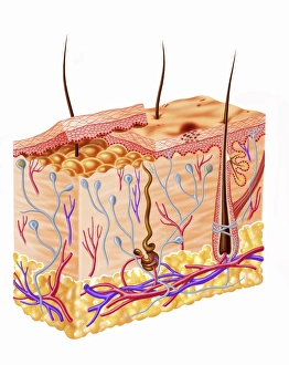
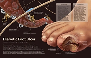
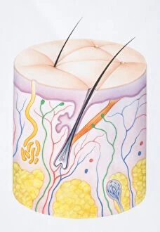
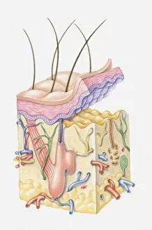
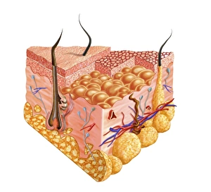
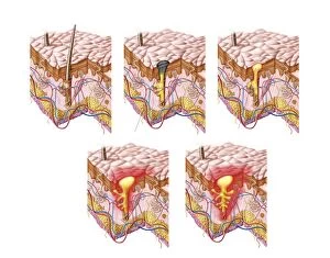
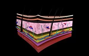
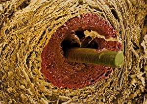
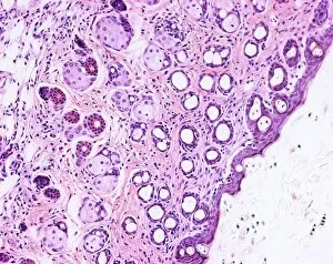
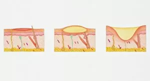
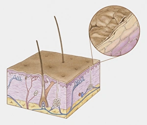
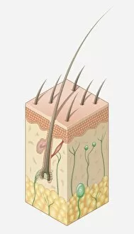
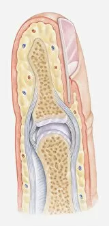
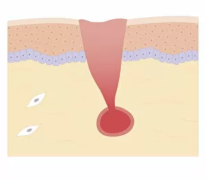
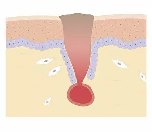
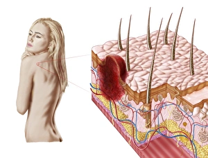
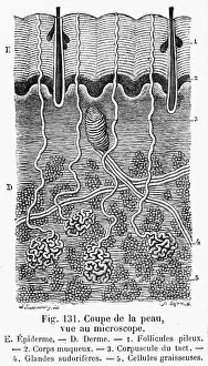
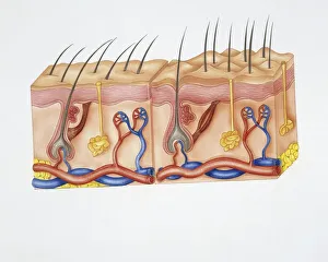
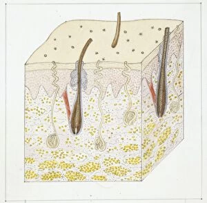
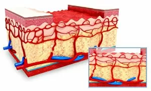
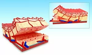
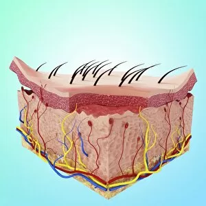
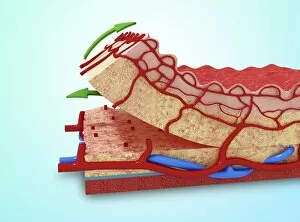
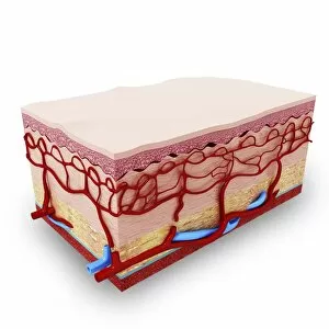
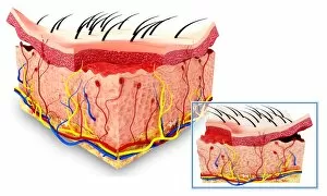
The dermis, a fascinating layer of our skin, plays a crucial role in protecting and maintaining our body. From skin disorders to intricate artwork, the dermis offers us an array of wonders to explore. In a SEM image of a section through human skin, we witness the complexity and beauty that lies beneath the surface. The layers of They can revealed, showcasing their unique functions and structures. Another illustration captures heat trapped by erect hairs in a cross-section of human skin. This phenomenon highlights how our body adapts to environmental changes and regulates temperature effectively. A colored SEM showcases blood vessels within the dermis, emphasizing its vital role in nourishing our skin cells. These intricate networks ensure proper circulation and contribute to overall health. Moving deeper into this microscopic world, we encounter various skin conditions depicted in cross-sections. A blackhead reveals clogged pores while papules showcase inflamed spots on the surface. Pustules demonstrate infected areas with pus accumulation, while whiteheads depict blocked follicles under the skin's surface. Delving further into anatomy, we explore how different layers interact harmoniously within the dermis. Each layer serves distinct purposes – from epidermis protection to collagen production – creating a resilient shield against external factors. Histopathology and pathophysiology come into play when examining diabetic foot ulcers' impact on the dermal layer. Understanding these processes helps medical professionals provide effective treatments for patients suffering from such conditions. Whether it is appreciating its artistic qualities or unraveling its complexities during medical research, exploring the wonders of the dermis unveils both aesthetic marvels and critical insights into human health.