
Pores Collection
"Pores: Unveiling the Hidden World" Pores, those tiny openings found in various natural and man-made structures, hold secrets waiting to be discovered


All Professionally Made to Order for Quick Shipping
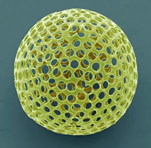
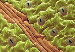
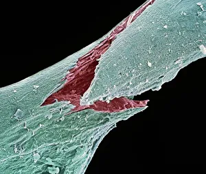
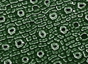
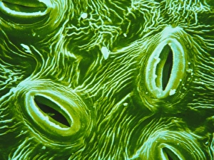
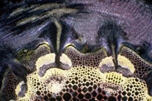
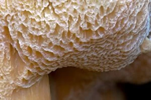
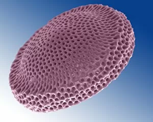
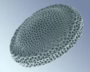
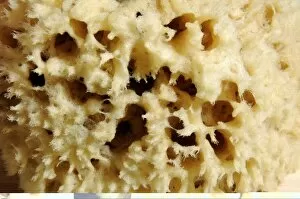
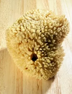
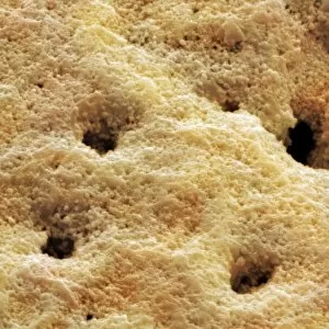
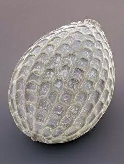
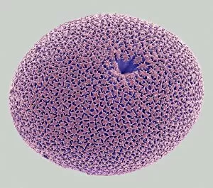
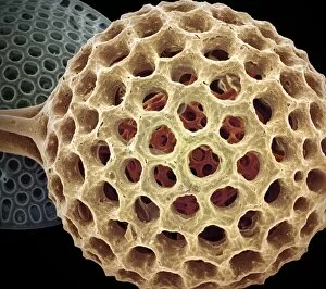
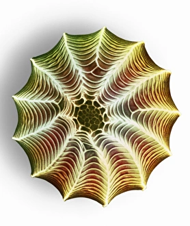
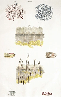
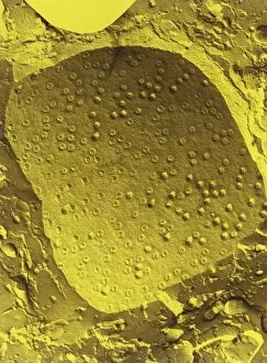
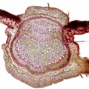
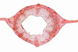
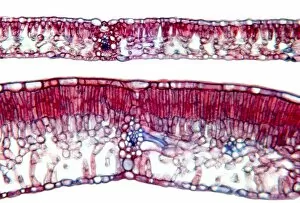
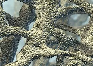
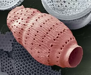
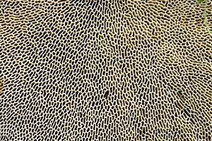
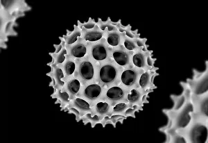
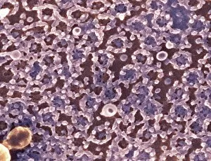
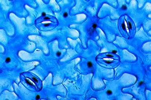
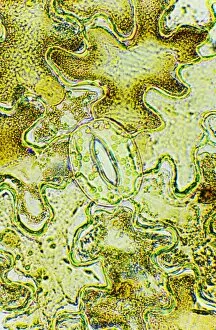
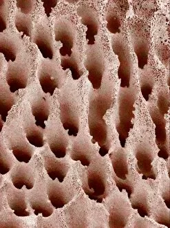
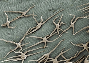
"Pores: Unveiling the Hidden World" Pores, those tiny openings found in various natural and man-made structures, hold secrets waiting to be discovered. From osteoporotic bone to leaf pores, these microscopic gateways offer a glimpse into the intricate workings of our world. In the realm of science, scanning electron microscopy (SEM) has allowed us to explore the fascinating intricacies of pores. Picture No. 11675628 reveals the delicate network of osteoporotic bone, highlighting how these minuscule channels contribute to its strength and resilience. Moving on from bones to nature's wonders, SEM captures the beauty of leaf pores or stomata on an Elder leaf. These minute structures play a vital role in regulating gas exchange for photosynthesis and transpiration in plants. Venturing deeper into unseen realms, we encounter Acrosphaera radiolarian under SEM. These marine microorganisms showcase their complex skeletal structure with countless intricate pores that aid in nutrient absorption and locomotion. Zooming even closer under SEM's watchful eye, we discover small white butterfly eggs revealing their secret through magnification. The detailed examination unravels unique pore patterns that serve as entry points for oxygen exchange during embryonic development. Shifting gears from nature's wonders to oceanic marvels, a close-up portrait captures a tiger shark gracefully gliding near Hawaii's surface. Its sleek skin bears evidence of tiny dermal denticles with minute pores acting as sensory receptors within this majestic predator's domain. Delving further underwater brings us face-to-face with another inhabitant - the Grey Reef Shark. Its textured skin showcases numerous specialized dermal denticles housing microscopic openings that enhance hydrodynamics while navigating vast ocean depths. Stepping away from living organisms but still exploring hidden worlds brings us to brown-rot fungi thriving at Clumber Park in Nottinghamshire. A close-up detail highlights their porous surfaces responsible for breaking down wood fibers—a crucial step in the natural recycling process.