Fraud Blocker Skip to main content

Pores Collection (page 2)

"Pores: Unveiling the Hidden World" Pores, those tiny openings found in various natural and man-made structures, hold secrets waiting to be discovered

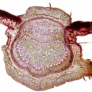
Background imagePores Collection: Beech tree leaf, light micrograph

Beech tree leaf, light micrograph
Beech tree leaf. Light micrograph of a section through the leaf of a common beech tree (Fagus sylvatica), showing the midrib

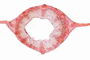
Background imagePores Collection: Sweet pea stem, light micrograph

Sweet pea stem, light micrograph
Sweet pea stem. Light micrograph of a section through the hollow stem of a sweet pea (Lathyrus odoratus) plant, showing a ring of vascular bundles

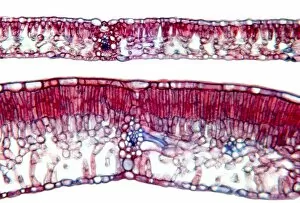
Background imagePores Collection: Beech tree leaves, light micrograph

Beech tree leaves, light micrograph
Beech tree leaves. Light micrograph of a section through two leaves from different parts of a common beech tree (Fagus sylvatica)

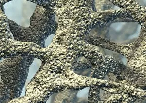
Background imagePores Collection: Osteoporotic bone, artwork

Osteoporotic bone, artwork
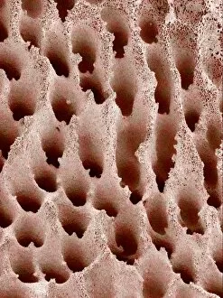
Osteoporosis. Computer artwork of osteoporotic bone. Osteoporosis (brittle bone disease) causes a reduction in overall bone mass and an increase in the bones porosity

Background imagePores Collection: Aquaporins, artwork

Aquaporins, artwork
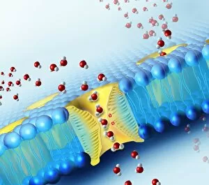
Aquaporins. Computer artwork of water molecules (red and white) passing through pores (yellow) in a cell membrane (blue). These pores are proteins known as aquaporins that are embedded in

Background imagePores Collection: Photosynthesis, diagram

Photosynthesis, diagram
Photosynthesis. Diagram showing the inputs and outputs (coloured arrows) involved in photosynthesis in the leaf of a plant

Background imagePores Collection: mRNA leaving the nucleus, artwork

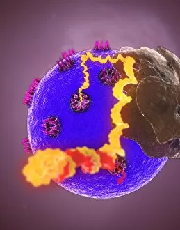
mRNA leaving the nucleus, artwork. mRNA (messenger ribonucleic acid, orange) is the intermediary molecule between DNA (deoxyribonucleic acid) and its protein product

Background imagePores Collection: Radiolarian, SEM

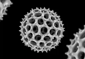
Radiolarian, SEM
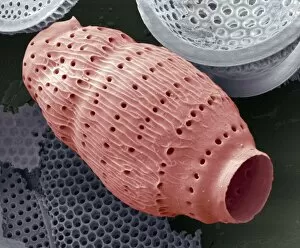
Radiolarian. Coloured scanning electron micrograph (SEM) of the shell of a radiolarian. Radiolaria are single-celled protozoans that are found in marine plankton

Background imagePores Collection: Bracket fungus (Daedaleopsis confragosa)

Bracket fungus (Daedaleopsis confragosa)
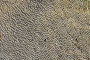
Blushing bracket fungus (Daedaleopsis confragosa). Close-up of the underside of the blushing bracket fungus showing its pores. The pores are the ends of the tubes within which the spores are produced

Background imagePores Collection: Radiolarian skeleton, artwork

Radiolarian skeleton, artwork
Radiolarian skeleton. Computer artwork of the skeleton (test) of a radiolarian. Radiolaria are single-celled protozoans that are found in marine plankton

Background imagePores Collection: Nuclear pore complexes, SEM

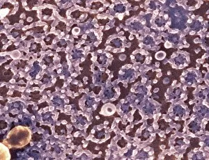
Nuclear pore complexes, SEM
Nuclear pore complexes. Coloured scanning electron micrograph (SEM) of the surface of a cell nucleus showing the numerous nuclear pore complexes (NPCs, rings) in its envelope

Background imagePores Collection: Blocked pore in acne, artwork

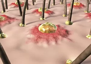
Blocked pore in acne, artwork
Blocked pore in acne, computer artwork. Sweat pore (centre) that has become blocked (centre) due to acne. Acne is a skin disorder in which the sebaceous glands of the skin become inflamed (red areas)

Background imagePores Collection: Plant stomata, light micrograph

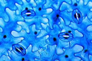
Plant stomata, light micrograph
Plant stomata. Light micrograph of stomatal pores on the surface of a kidney bean (Phaseolus sp.) leaf. The stomata are gaps (white) within two guard cells (blue, kidney-shaped)

Background imagePores Collection: Tea leaf, light micrograph

Tea leaf, light micrograph
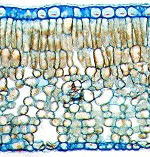
Tea leaf. Light micrograph of a cross-section through a tea (Camellia sinensis) leaf. The upper and lower epidermis on the surfaces of the leaf are blue

Background imagePores Collection: Plant stoma, light micrograph

Plant stoma, light micrograph
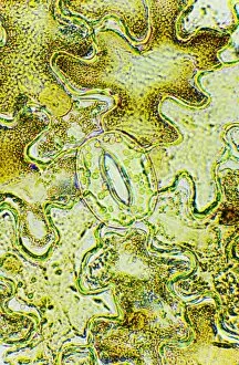
Plant stoma. Light micrograph of a stomatal pore (centre) on the surface of a stinging nettle (Urtica dioica) leaf. The stomata are gaps (white) within two guard cells (kidney-shaped)

Background imagePores Collection: Mushroom surface, SEM

Mushroom surface, SEM
Mushroom surface. Coloured scanning electron micrograph (SEM) of the spore-bearing surface of a hen of the woods mushroom (Grifola frondosa). This surface is found on the underside of the mushroom

Background imagePores Collection: Ivy stem, SEM

Ivy stem, SEM
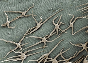
Ivy stem. Coloured scanning electron micrograph (SEM) of the surface of an ivy plant stem. The star- shaped trichomes, a type of modified hair cell, may function in preventing water loss

Background imagePores Collection: Section through leaf of Zinnia

Section through leaf of Zinnia
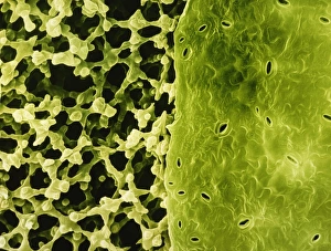
False-colour scanning electron micrograph of the under surface of a leaf, Zinnia elegans, showing a part of the epidermis removed to reveal the spongy mesophyll layer (left) under the surface

Background imagePores Collection: Sunflower leaf, SEM

Sunflower leaf, SEM
Sunflower leaf. Coloured scanning electron micrograph (SEM) of the underside of a sunflower leaf (Helianthus annuus). The green and yellow structures are trichomes

Background imagePores Collection: Open stomata, SEM

Open stomata, SEM
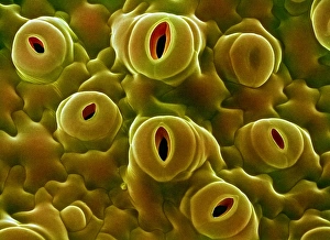
Open stomata. Coloured scanning electron micrograph (SEM) of open stomata on the surface of a tobacco leaf (Nicotiana tabacum)

Background imagePores Collection: Tomato leaf surface, SEM

Tomato leaf surface, SEM
Tomato leaf surface. Coloured scanning electron micrograph (SEM) of the surface of a tomato leaf (Lycopersicon esculentum). It is covered in trichomes (red), which secrete oil, and hairs

Background imagePores Collection: Daisy leaf surface, SEM

Daisy leaf surface, SEM
Daisy leaf. Coloured scanning electron micrograph (SEM) of the surface of a daisy leaf (family Asteraceae, formerly Compositae). Three stomata (leaf pores) can be seen

Background imagePores Collection: Plant pores, SEM

Plant pores, SEM
Plant pores. Coloured scanning electron micrograph (SEM) of stomata (pores) on the pistil of a bellflower (Adenophora sp.). Stomata are the site of gaseous exchange, or respiration

Background imagePores Collection: Open stoma, SEM

Open stoma, SEM
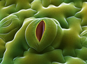
Open stoma. Coloured scanning electron micrograph (SEM) of an open stoma on the surface of a tobacco leaf (Nicotiana tabacum)

Background imagePores Collection: Caterpillar spiracles, light micrograph

Caterpillar spiracles, light micrograph
Caterpillar spiracles. Light micrograph of part of the skin of a caterpillar showing part of the tracheal system for bringing oxygen to the body tissues

Background imagePores Collection: Caterpillar skin, light micrograph

Caterpillar skin, light micrograph. This section of part of the skin shows two half segments. The skin is covered in small hairs or setae (black), and brown structures called tubercles and verrucae

Background imagePores Collection: Caterpillar spiracle, light micrograph

Caterpillar spiracle, light micrograph
Caterpillar spiracle. Polarised light micrograph of part of the skin of a caterpillar, showing an air hole (spiracle, dark oval, lower right) leading to a breathing tube (trachea)

Background imagePores Collection: Zeolite A crystal structure

Zeolite A crystal structure
Zeolite A crystal, molecular model. Zeolite A is a synthetic alumino-silicate material manufactured for use as a molecular sieve


All Professionally Made to Order for Quick Shipping

"Pores: Unveiling the Hidden World" Pores, those tiny openings found in various natural and man-made structures, hold secrets waiting to be discovered. From osteoporotic bone to leaf pores, these microscopic gateways offer a glimpse into the intricate workings of our world. In the realm of science, scanning electron microscopy (SEM) has allowed us to explore the fascinating intricacies of pores. Picture No. 11675628 reveals the delicate network of osteoporotic bone, highlighting how these minuscule channels contribute to its strength and resilience. Moving on from bones to nature's wonders, SEM captures the beauty of leaf pores or stomata on an Elder leaf. These minute structures play a vital role in regulating gas exchange for photosynthesis and transpiration in plants. Venturing deeper into unseen realms, we encounter Acrosphaera radiolarian under SEM. These marine microorganisms showcase their complex skeletal structure with countless intricate pores that aid in nutrient absorption and locomotion. Zooming even closer under SEM's watchful eye, we discover small white butterfly eggs revealing their secret through magnification. The detailed examination unravels unique pore patterns that serve as entry points for oxygen exchange during embryonic development. Shifting gears from nature's wonders to oceanic marvels, a close-up portrait captures a tiger shark gracefully gliding near Hawaii's surface. Its sleek skin bears evidence of tiny dermal denticles with minute pores acting as sensory receptors within this majestic predator's domain. Delving further underwater brings us face-to-face with another inhabitant - the Grey Reef Shark. Its textured skin showcases numerous specialized dermal denticles housing microscopic openings that enhance hydrodynamics while navigating vast ocean depths. Stepping away from living organisms but still exploring hidden worlds brings us to brown-rot fungi thriving at Clumber Park in Nottinghamshire. A close-up detail highlights their porous surfaces responsible for breaking down wood fibers—a crucial step in the natural recycling process.