Beech tree leaves, light micrograph

![]()
Wall Art and Photo Gifts from Science Photo Library
Beech tree leaves, light micrograph
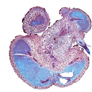
Beech tree leaves. Light micrograph of a section through two leaves from different parts of a common beech tree (Fagus sylvatica). Some leaves of a beech tree get the maximum exposure to sunlight whilst others get less sunlight because they are shaded; e.g. other leaves are above them. Therefore the leaves vary in their structure according to how much sunlight they receive. The lamina of a sun leaf (lower) is thicker; it has two layers of palisade mesophyll tissue and more chloroplasts. It also has a deeper spongy mesophyll layer with more chloroplasts and storage for starch, more air space for gases to get to the cells, bigger veins and xylem vessels to receive water and minerals, and bigger phloem vessels to carry food around the plant. The shade leaf
Science Photo Library features Science and Medical images including photos and illustrations
Media ID 6338513
© DR KEITH WHEELER/SCIENCE PHOTO LIBRARY
Bundles Cell Biology Cell Wall Common Beech Compared Comparing Comparison Cytological Cytology Dicot Dicots Dicotyledon Dicotyledons Difference Differences Epidermis Fagus Sylvatica Ground Tissue Histological Histology Lamina Layer Layers Microscopy Mid Rib Palisade Mesophyll Parenchyma Phloem Pore Pores Sclerenchyma Spongy Mesophyll Stain Stained Stomata Structural Structures Supporting Supportive Tissue Tissue Vascular Bundle Xylem Cells Light Micrograph Light Microscope Section Sectioned
EDITORS COMMENTS
This print showcases the intricate details of beech tree leaves, as seen under a light microscope. The image reveals two leaves from different parts of a common beech tree (Fagus sylvatica), highlighting how their structures vary due to differences in sunlight exposure. Some leaves are fortunate enough to receive maximum sunlight, while others are shaded by neighboring foliage. The sun leaf, depicted on the lower side of the micrograph, exhibits a thicker lamina with two layers of palisade mesophyll tissue and an abundance of chloroplasts. Its spongy mesophyll layer is deeper and also contains more chloroplasts for photosynthesis. Additionally, this leaf possesses larger veins and xylem vessels that transport water and minerals, as well as bigger phloem vessels responsible for distributing food throughout the plant. In contrast, the shade leaf demonstrates distinct differences in its structure compared to its sunlit counterpart. This variation arises from receiving less direct sunlight. While both types of leaves serve their purpose within the plant's ecosystem, they have adapted differently based on their individual needs. Through this stunning micrograph captured by Science Photo Library, we gain insight into the fascinating world of botany and appreciate nature's ability to adapt and thrive under varying conditions.
MADE IN THE USA
Safe Shipping with 30 Day Money Back Guarantee
FREE PERSONALISATION*
We are proud to offer a range of customisation features including Personalised Captions, Color Filters and Picture Zoom Tools
SECURE PAYMENTS
We happily accept a wide range of payment options so you can pay for the things you need in the way that is most convenient for you
* Options may vary by product and licensing agreement. Zoomed Pictures can be adjusted in the Cart.