Fraud Blocker Skip to main content

Sclerenchyma Collection

Sclerenchyma, a type of plant tissue, is known for its strength and support

Background imageSclerenchyma Collection: Balsa wood structure, SEM

Balsa wood structure, SEM
Balsa wood structure, coloured scanning electron micrograph (SEM). This wood is from the balsa tree (Ochroma pyramidale). This is a longitudinal section through the wood

Background imageSclerenchyma Collection: Ammophila arenaria leaf, light micrograph

Ammophila arenaria leaf, light micrograph
Ammophila arenaria leaf. Polarised light micrograph of a section through a marram grass (Ammophila arenaria) leaf, showing the characteristics that help reduce water loss

Background imageSclerenchyma Collection: Beech tree leaf, light micrograph

Beech tree leaf, light micrograph
Beech tree leaf. Light micrograph of a section through the leaf of a common beech tree (Fagus sylvatica), showing the midrib

Background imageSclerenchyma Collection: Sycamore leaf vein, light micrograph

Sycamore leaf vein, light micrograph
Sycamore leaf vein. Light micrograph of a section through the midrib (vein) of a leaf from a sycamore (Acer pseudoplatanus) tree

Background imageSclerenchyma Collection: Geranium stem, light micrograph

Geranium stem, light micrograph
Geranium stem. Light micrograph of a section through a young stem of a geranium (Pelargonium sp.) plant. The outer stem is covered with a thin epidermis (red) which has stomata

Background imageSclerenchyma Collection: Sweet pea stem, light micrograph

Sweet pea stem, light micrograph
Sweet pea stem. Light micrograph of a section through the hollow stem of a sweet pea (Lathyrus odoratus) plant, showing a ring of vascular bundles

Background imageSclerenchyma Collection: Beech tree leaves, light micrograph

Beech tree leaves, light micrograph
Beech tree leaves. Light micrograph of a section through two leaves from different parts of a common beech tree (Fagus sylvatica)

Background imageSclerenchyma Collection: Fern stem, light micrograph

Fern stem, light micrograph. Transverse section through a rachis (stem) of the bracken fern (Pteridium aquilinum). Under the outer epidermis (black) is a thin cortex (deep red)

Background imageSclerenchyma Collection: Horsetail stem, light micrograph

Horsetail stem, light micrograph. Transverse section through the stem of the common horsetail (Equisetum arvensis). The stem consists of an outer epidermis and an outer cortex of sclerenchyma cells

Background imageSclerenchyma Collection: Fern rhizome, light micrograph

Fern rhizome, light micrograph. Transverse section through the center of a rhizome from the Killarney fern (Trichomanes speciosum)

Background imageSclerenchyma Collection: Clubmoss stem, light micrograph

Clubmoss stem, light micrograph
Clubmoss stem, polarised light micrograph. Transverse section through the stem of the clubmoss Lycopodium clavatum. This is the central portion of the stem consisting of the inner cortex (red)

Background imageSclerenchyma Collection: Common broom stem, light micrograph

Common broom stem, light micrograph
Common broom stem. Light micrograph of a transverse section through the stem of a common broom (Salicornia europaea) plant

Background imageSclerenchyma Collection: Dutchmans pipe plant stem

Dutchmans pipe plant stem. Light micrograph of a section through the stem of a dutchmans pipe plant (Aristolochia sipho). The inner blue circle is a layer of sclerenchyma fibres

Background imageSclerenchyma Collection: Grass stem, SEM

Grass stem, SEM
Grass stem. Coloured scanning electron micrograph (SEM) of a section through a grass stem (family Graminaceae). At upper centre is the vascular bundle

Background imageSclerenchyma Collection: Mistletoe vascular bundle, LM

Mistletoe vascular bundle, LM
Mistletoe vascular bundle. Light micrograph (LM) of a section of mistletoe (Viscum album) stem in cross-section, showing a vascular bundle

Background imageSclerenchyma Collection: Mistletoe stem, LM

Mistletoe stem, LM
Mistletoe stem. Light micrograph (LM) of a mistletoe (Viscum album) stem in cross-section, showing nine vascular bundles radiating out from the centre of the stem

Background imageSclerenchyma Collection: She-oak stem, light micrograph

She-oak stem, light micrograph
She-oak stem. Light micrograph of a transverse section through the stem of the Australian she-oak (Casuarina equisetifolia). The she-oak is a drought plant (xerophyte)

Background imageSclerenchyma Collection: Clematis stem, light micrograph

Clematis stem, light micrograph
Clematis stem. Light micrograph of a transverse section through the stem of a clematis (Clematis flammula) plant. At the centre of the stem is a large area of pith, consisting of parenchyma cells

Background imageSclerenchyma Collection: Liana stem, light micrograph

Liana stem, light micrograph
Liana stem. Light micrograph of a transverse section through the stem of a liana (Aristolochia tormentosa), or woody vine. At the centre of the stem is the pith, consisting of parenchyma cells

Background imageSclerenchyma Collection: Light Micrograph (LM) of a transverse section showing Sclerenchyma Ground Tissue in Helianthus stem

Light Micrograph (LM) of a transverse section showing Sclerenchyma Ground Tissue in Helianthus stem, magnification x 600


All Professionally Made to Order for Quick Shipping

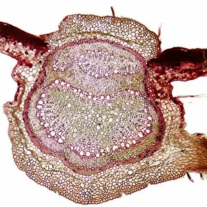
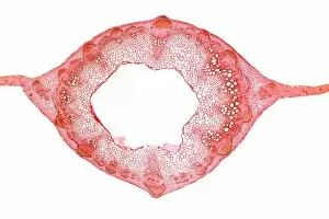
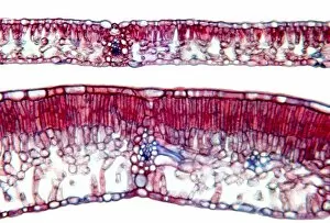
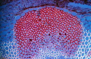
Sclerenchyma, a type of plant tissue, is known for its strength and support. Its structure can be compared to that of balsa wood when observed under a scanning electron microscope (SEM). The SEM reveals intricate patterns resembling the lightweight yet sturdy composition of balsa wood. In addition to its resemblance to balsa wood they can also be found in various other plant structures. For instance, in maize stems, light micrographs show the presence cells providing rigidity and structural integrity. These cells are arranged in bundles or strands throughout the stem, reinforcing it against bending or breaking. Another example is seen in Ammophila arenaria leaves. Multiple light micrographs capture the intricate network cells within these leaves. This arrangement helps maintain their shape even under harsh conditions such as strong winds or shifting sand dunes where Ammophila arenaria thrives. The repetitive appearance of Ammophila arenaria leaf micrographs emphasizes how vital sclerenchyma is for this particular species' survival. It highlights nature's ingenious design by incorporating this tissue into their leaves repeatedly to withstand environmental challenges. Maize stems also exhibit similar adaptations with multiple light micrograph images showcasing the presence cells throughout their structure. These cells provide mechanical support and contribute to overall stem strength necessary for upright growth and resistance against external forces. Furthermore, clubmoss stems display a comparable pattern when examined using light microscopy techniques. Sclerenchyma plays an essential role here too by enhancing stability and preventing collapse during growth stages. Overall, these visual representations emphasize the importance and versatility tissue across different plants' anatomies. Whether it resembles balsa wood under SEM or appears as interconnected networks within maize stems, Ammophila arenaria leaves, or clubmoss stems through light microscopy - one thing remains clear.