Fine Art Print : Small intestine structures, artwork

![]()
Fine Art Prints From Science Photo Library
Small intestine structures, artwork
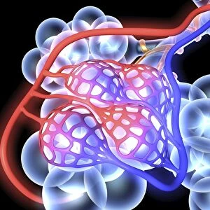
Small intestine structures. Artwork sequence of magnified views of the small intestine, with the magnification increasing from top to bottom. At top is a cutaway view, showing the lumen along which food passes as it is being digested. The three main layers (red, smooth muscle; orange, submucosa; yellow, mucosa) are shown here and in the second image, which shows large folds on the intestinal wall on which are projections called villi. Three sectioned villi are in the third image, with blood vessels (red and blue) and lymph vessels (green). The fourth image shows the enterocyte cells (microvilli brush border) that absorb nutrients
Science Photo Library features Science and Medical images including photos and illustrations
Media ID 6328409
© ART FOR SCIENCE/SCIENCE PHOTO LIBRARY
Alimentary Canal Arterial Brush Border Capillaries Capillary Cell Biology Cut Away Diagram Digestion Digestive System Enterocyte Enterocytes Epithelium Fold Folds Gastroenterology Histological Histology Inset Intestinal Intestine Lumen Lymph Vessel Lymphatic System Magnified Microvilli Microvillus Mucosa Series Small Intestine Small Intestines Smooth Muscle Sub Mucosa Venous Vessels Villi Villus Artery Cells Vein
20"x20" (+3" Border) Fine Art Print
Discover the intricate beauty of the human body with our Fine Art Prints from Media Storehouse, featuring the captivating artwork "Small Intestine Structures" by Science Photo Library. This mesmerizing sequence showcases magnified views of the small intestine, revealing its complex labyrinthine network. Each print offers a unique perspective, with magnification increasing from top to bottom. Bring the wonders of science into your home or office and ignite curiosity with these stunning, high-quality Fine Art Prints.
21x7 image printed on 27x13 Fine Art Rag Paper with 3" (76mm) white border. Our Fine Art Prints are printed on 300gsm 100% acid free, PH neutral paper with archival properties. This printing method is used by museums and art collections to exhibit photographs and art reproductions.
Our fine art prints are high-quality prints made using a paper called Photo Rag. This 100% cotton rag fibre paper is known for its exceptional image sharpness, rich colors, and high level of detail, making it a popular choice for professional photographers and artists. Photo rag paper is our clear recommendation for a fine art paper print. If you can afford to spend more on a higher quality paper, then Photo Rag is our clear recommendation for a fine art paper print.
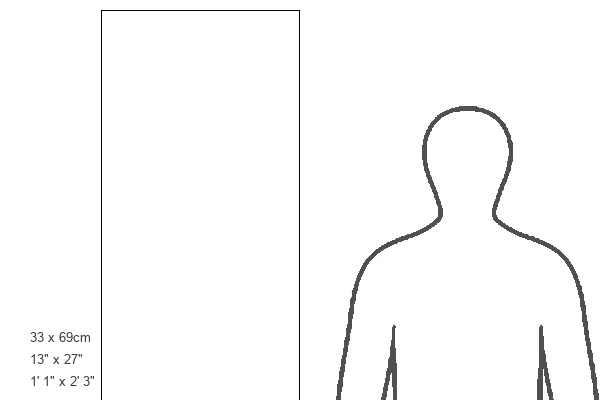
Estimated Image Size (if not cropped) is 17.7cm x 44.6cm (7" x 17.6")
Estimated Product Size is 33cm x 68.6cm (13" x 27")
These are individually made so all sizes are approximate
Artwork printed orientated as per the preview above, with portrait (vertical) orientation to match the source image.
EDITORS COMMENTS
This artwork sequence titled "Small Intestine Structures" takes us on a mesmerizing journey through the intricate anatomy of the small intestine. The print showcases a series of magnified views, each revealing a deeper layer of this vital organ. Starting at the top, we are presented with a cutaway view that exposes the lumen, where food passes during digestion. As our eyes descend, we encounter three distinct layers: smooth muscle in vibrant red, submucosa in striking orange, and mucosa in radiant yellow. These layers play crucial roles in ensuring proper digestion and nutrient absorption. Moving further down, large folds adorn the intestinal wall like majestic landscapes. Upon these folds reside tiny projections called villi - highlighted beautifully in the second image. It is within these delicate structures that nutrient absorption truly occurs. The third image provides an even closer look at three sectioned villi accompanied by blood vessels painted vividly in red and blue and lymph vessels depicted as refreshing green streams. This portrayal emphasizes their essential role in transporting nutrients throughout our body. Finally, we are introduced to enterocyte cells adorned with microvilli brush borders - showcased elegantly in the fourth image. These remarkable cells possess extraordinary abilities to absorb nutrients efficiently. Through this stunning artwork sequence created by Science Photo Library, viewers gain an appreciation for both the complexity and beauty found within our own bodies' digestive system – reminding us once again of nature's incredible design.
MADE IN THE USA
Safe Shipping with 30 Day Money Back Guarantee
FREE PERSONALISATION*
We are proud to offer a range of customisation features including Personalised Captions, Color Filters and Picture Zoom Tools
SECURE PAYMENTS
We happily accept a wide range of payment options so you can pay for the things you need in the way that is most convenient for you
* Options may vary by product and licensing agreement. Zoomed Pictures can be adjusted in the Basket.