Fine Art Print : Paprosky femur defect, type IIIA lateral

![]()
Fine Art Prints From Science Photo Library
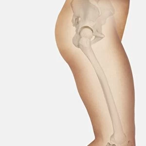
Paprosky femur defect, type IIIA lateral
Paprosky femur defect. Cutaway artwork of bone degradation in a type IV medial-lateral femur cortex defect (Paprosky classification system). This system is used for revision (replacement or repair) of a hip implant. The ball and socket part of the implant is at top. The rest of the implant consists of a shaft inside the femur (thigh bone). The amount of degradation in the cortical (outer) bone layer determines the amount of bone grafting needed, and whether a cementless implant can replace a cemented one. Type IV defects make the shaft unable to support weight (see C016/6620 for other types). Named for US surgeon Wayne G. Paprosky, the classification was developed in the 1980s and 1990s
Science Photo Library features Science and Medical images including photos and illustrations
Media ID 9214781
© D & L GRAPHICS / SCIENCE PHOTO LIBRARY
Arthrology Arthroplasty Bone Cement Cemented Cortex Cortical Cutaway Defect Defects Diagram Diaphysis Femur Grafting Hip Implant Hip Replacement Hip Revision Joint Medial Orthopaedic Orthopaedics Orthopedic Orthopedics Osteological Osteology Prostheses Prosthesis Prosthetic Prosthetics Repair Replacement Shaft Surgery Surgical Type Types Condition Cutouts Disorder Type 3
21"x14" (+3" Border) Fine Art Print
Discover the intricacies of human anatomy with Media Storehouse's Fine Art Prints. This captivating image, titled "Paprosky Femur Defect, Type IIIA Lateral," is a meticulously crafted cutaway artwork by D & L Graphics / Science Photo Library. It provides a mesmerizing insight into the complexities of bone degradation in the medial-lateral femur cortex defect, as classified by Paprosky. Ideal for medical professionals, educators, or anyone with a deep fascination for anatomy, these Fine Art Prints bring scientific knowledge to life with their exceptional detail and visual clarity.
21x14 image printed on 27x20 Fine Art Rag Paper with 3" (76mm) white border. Our Fine Art Prints are printed on 300gsm 100% acid free, PH neutral paper with archival properties. This printing method is used by museums and art collections to exhibit photographs and art reproductions.
Our fine art prints are high-quality prints made using a paper called Photo Rag. This 100% cotton rag fibre paper is known for its exceptional image sharpness, rich colors, and high level of detail, making it a popular choice for professional photographers and artists. Photo rag paper is our clear recommendation for a fine art paper print. If you can afford to spend more on a higher quality paper, then Photo Rag is our clear recommendation for a fine art paper print.
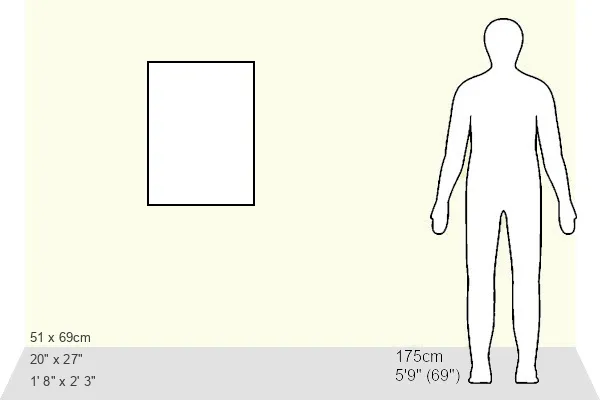
Estimated Image Size (if not cropped) is 28.4cm x 53.3cm (11.2" x 21")
Estimated Product Size is 50.8cm x 68.6cm (20" x 27")
These are individually made so all sizes are approximate
Artwork printed orientated as per the preview above, with portrait (vertical) orientation to match the source image.
EDITORS COMMENTS
This detailed print showcases a Paprosky femur defect, specifically a type IIIA lateral defect. The image is an intricate cutaway artwork that depicts the degradation of bone in the medial-lateral femur cortex, following the Paprosky classification system. This classification system is crucial for guiding revision procedures of hip implants. At the top of the illustration, we can observe the ball and socket component of the implant, while the rest consists of a shaft placed inside the femur (thigh bone). The severity of cortical bone degradation determines whether bone grafting is necessary and if a cementless implant can replace a cemented one. Type IV defects render the shaft incapable of supporting weight. Named after esteemed US surgeon Wayne G. Paprosky, this classification was developed during the 1980s and 1990s to aid orthopedic surgeons in determining appropriate treatment plans for patients requiring hip implant revisions. The image provides valuable insight into this medical condition by highlighting various anatomical structures such as metaphysis, metaphyseal region, diaphysis, and cortical layers affected by deterioration. It serves as an educational tool for healthcare professionals involved in arthroplasty surgeries or those studying osteology and arthrology. With its white background and precise detailing, this print from D & L GRAPHICS / SCIENCE PHOTO LIBRARY offers an informative visual representation that aids understanding of large bone defects like Paprosky femur defects within medical contexts.
MADE IN THE USA
Safe Shipping with 30 Day Money Back Guarantee
FREE PERSONALISATION*
We are proud to offer a range of customisation features including Personalised Captions, Color Filters and Picture Zoom Tools
SECURE PAYMENTS
We happily accept a wide range of payment options so you can pay for the things you need in the way that is most convenient for you
* Options may vary by product and licensing agreement. Zoomed Pictures can be adjusted in the Basket.