Home > Science > SEM
Myelinated nerves, SEM C013 / 7138

![]()
Wall Art and Photo Gifts from Science Photo Library
Myelinated nerves, SEM C013 / 7138
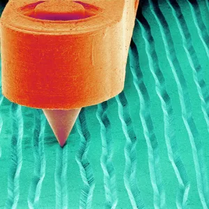
Myelinated nerves. Coloured scanning electron micrograph (SEM) of a section through a myelinated nerve fibre (axon, beige, centre) from the sciatic nerve. Myelin (orange and green) is an insulating fatty layer that surrounds the nerve fibre, increasing the speed at which nerve impulses travel. It is formed when Schwann cells wrap around the fibre, depositing layers of myelin between each coil. The outermost layer (green) consists of the Schwann cells cytoplasm and is known as the neurolemma or sheath of Schwann. Magnification: x8000, when printed 10 centimetres wide
Science Photo Library features Science and Medical images including photos and illustrations
Media ID 9198683
© STEVE GSCHMEISSNER/SCIENCE PHOTO LIBRARY
Axon Colored Cytoplasm Endoneurium Fatty Fibre Fibres Insulated Insulating Insulation Myelin Myelinated Nerve Neurolemma Neuron Neurone Neurones Neurons Neuroscience Phospholipid Schwann Cell Sheath Sheath Of Schwann Sheathed System Cells Nervous System Neurological Neurology Section Sectioned
EDITORS COMMENTS
This print showcases the intricate beauty of myelinated nerves, providing a glimpse into the complex world of our nervous system. In this coloured scanning electron micrograph (SEM), we are presented with a section through a myelinated nerve fibre from the sciatic nerve. The beige axon takes center stage, surrounded by an insulating fatty layer known as myelin, depicted in vibrant shades of orange and green. Myelin plays a crucial role in enhancing the speed at which nerve impulses travel along these fibres. It is formed when Schwann cells wrap around the fibre, depositing layers of myelin between each coil. The outermost layer, represented by the striking green coloration, consists of Schwann cell cytoplasm and is referred to as the neurolemma or sheath of Schwann. At a magnification level of x8000 when printed 10 centimetres wide, this image allows us to appreciate the remarkable details that make up our biological systems. From its vivid colors to its intricate structure, this SEM offers insights into both healthy anatomy and neurological function. Photographed by Steve Gschmeissner for Science Photo Library, this mesmerizing image invites us to marvel at nature's complexity while deepening our understanding of how neurons communicate within our bodies.
MADE IN THE USA
Safe Shipping with 30 Day Money Back Guarantee
FREE PERSONALISATION*
We are proud to offer a range of customisation features including Personalised Captions, Color Filters and Picture Zoom Tools
SECURE PAYMENTS
We happily accept a wide range of payment options so you can pay for the things you need in the way that is most convenient for you
* Options may vary by product and licensing agreement. Zoomed Pictures can be adjusted in the Cart.