Home > Popular Themes > Human Body
Brain fibres, DTI MRI scan C017 / 7038

![]()
Wall Art and Photo Gifts from Science Photo Library
Brain fibres, DTI MRI scan C017 / 7038
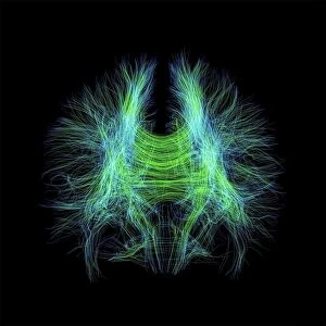
Brain fibres. 3D diffusion tensor imaging (DTI) magnetic resonance imaging (MRI) scan of a selection of nerve pathways (green/yellow) in the brain. The brain is seen from above, with the front of the brain at bottom. The ventricles are pink and the brains cortical surface (only shown at left) is blue. Diffusion tensor imaging measures the direction of water diffusion, which in the brain reveals the orientation of nerve fibres. The technique is also known as tractography, with the resulting image known as a tractogram
Science Photo Library features Science and Medical images including photos and illustrations
Media ID 9270495
© SHERBROOKE CONNECTIVITY IMAGING LAB/SCIENCE PHOTO LIBRARY
Brain Imaging Brain Scan Central Nervous System Cerebral Cerebrum Diffusion Tensor Imaging Dti Scan Fiber Fibers Fibre Fibres Imaging Technique Magnetic Resonance Imaging Mri Scan Mri Scanner Nerve Nerve Fibre Nerves Neural Pathway Neural Tract Paths Pathway Pathways Structural Tractogram Tractography Ventricle Ventricles White Matter Brain Neurological Neurology
EDITORS COMMENTS
This print showcases the intricate network of brain fibres, captured through a cutting-edge imaging technique called diffusion tensor imaging (DTI) magnetic resonance imaging (MRI). The image reveals a mesmerizing array of nerve pathways depicted in vibrant green and yellow hues against a striking black background. Viewed from above, with the front of the brain at the bottom, this visual representation offers an unprecedented glimpse into the complexity and beauty of our neural architecture. The pink ventricles serve as focal points within this composition, while on the left side, we catch a glimpse of the brain's cortical surface in serene blue tones. DTI measures water diffusion direction to unveil nerve fibre orientation within our brains. This revolutionary technique is also known as tractography, with resulting images referred to as tractograms. As we delve into this photograph's depths, it becomes evident that it represents far more than just an aesthetically pleasing arrangement. It symbolizes breakthroughs in medicine and biology by providing invaluable insights into healthy anatomical structures and neurological processes. By studying these neural pathways meticulously mapped out before us, scientists gain crucial knowledge about how our central nervous system functions. In essence, this remarkable image serves as a testament to human ingenuity and our relentless pursuit of understanding one of nature's most enigmatic creations – the human brain.
MADE IN THE USA
Safe Shipping with 30 Day Money Back Guarantee
FREE PERSONALISATION*
We are proud to offer a range of customisation features including Personalised Captions, Color Filters and Picture Zoom Tools
SECURE PAYMENTS
We happily accept a wide range of payment options so you can pay for the things you need in the way that is most convenient for you
* Options may vary by product and licensing agreement. Zoomed Pictures can be adjusted in the Cart.