
Brain Imaging Collection
"Unlocking the Secrets of the Brain: A Journey through Brain Imaging" Step into the fascinating world of brain imaging


All Professionally Made to Order for Quick Shipping
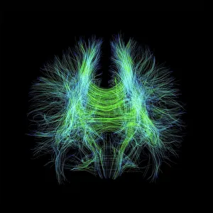
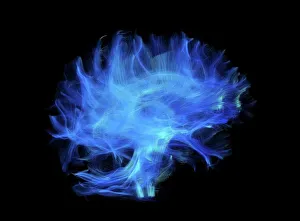
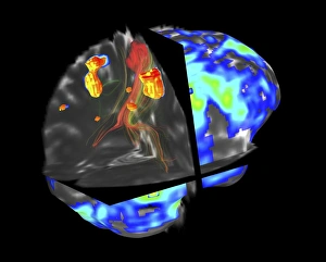
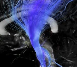
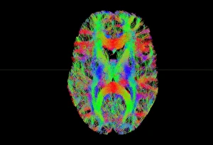
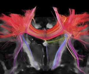
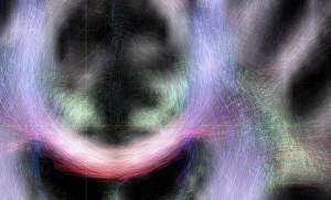
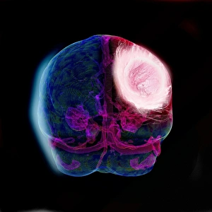
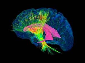
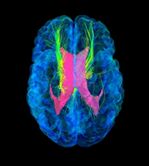
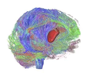
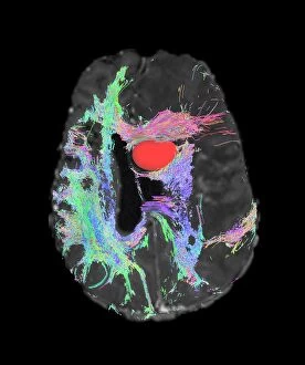
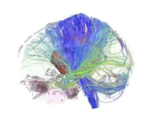
"Unlocking the Secrets of the Brain: A Journey through Brain Imaging" Step into the fascinating world of brain imaging, where cutting-edge technology allows us to explore the intricate network of brain fibres. With DTI MRI scans like C017/7099 and C017/7035, we delve deep into understanding these complex pathways that connect different regions of our brains. In our quest for knowledge, we encounter challenges such as brain tumours. However, with fMRI and tractography techniques like in scan C017/7102, we can visualize these tumors and their impact on neural connections. The corticospinal tract is no exception; its importance becomes evident through DTI MRI scan C017/7046. Tract density imaging reveals further insights into brain fibres (C017/7039), while DTI MRI scans shed light on the white matter within our brains. These invaluable tools aid in diagnosing conditions like glioblastoma brain tumours (C017/7048, C017/7055, and C017/7056). By utilizing advanced DTI modelling techniques (C017/7060), medical professionals gain a deeper understanding of these aggressive tumours' behavior. Amongst this vast sea of information lies the corpus callosum – a bridge connecting both hemispheres. Its significance unravels through DTI MRI scan C1074; it highlights how disruptions in this structure can affect communication between different parts of our brains. Brain imaging opens doors to new possibilities for diagnosis and treatment by unraveling mysteries hidden within our most vital organ. As technology continues to advance, so does our ability to comprehend the complexities that make each individual's mind unique.