
White Matter Collection
White matter refers to the intricate network of brain fibres that connect different regions of the brain, allowing for efficient communication and information processing


All Professionally Made to Order for Quick Shipping
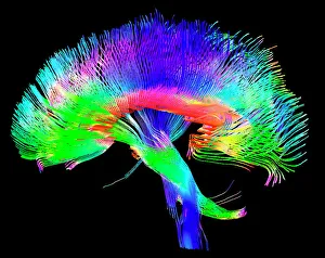
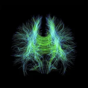
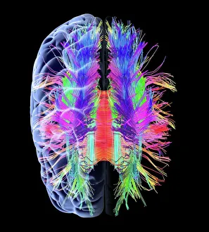
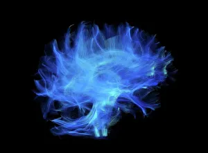
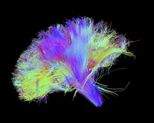
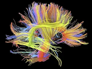
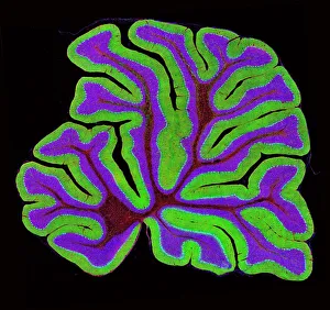
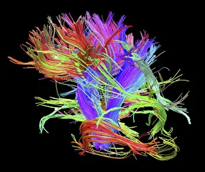
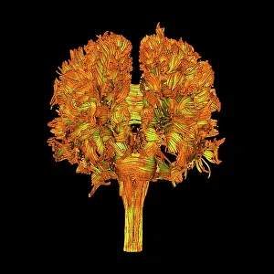
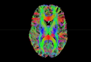
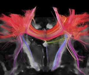
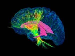
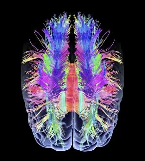
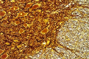
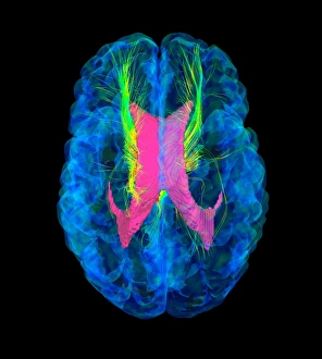
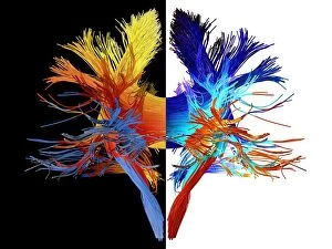
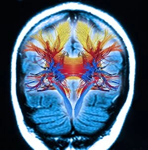
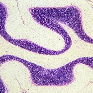
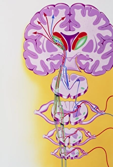
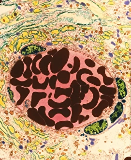
White matter refers to the intricate network of brain fibres that connect different regions of the brain, allowing for efficient communication and information processing. These pathways can be visualized using advanced imaging techniques such as DTI MRI scans. In one captivating artwork, titled "White Matter Fibres and Brain, " we witness a mesmerizing depiction of these neural connections. The piece showcases the complexity and beauty of our brain's white matter, highlighting its crucial role in cognitive function. Through DTI MRI scans like C017 / 7035 and C017 / 7099, scientists have been able to map out these intricate brain fibres with remarkable precision. These images provide valuable insights into how information flows through various regions of the brain, enabling us to better understand neurological disorders and develop targeted treatments. The importance cannot be overstated; it serves as a vital highway for transmitting electrical signals between different parts of our brains. Just like roads connecting cities, these pathways allow for efficient communication among diverse areas responsible for functions such as memory, language processing, and motor skills. One particularly intriguing image showcases the white matter fibres within the human cerebellum structure captured under a light microscope. This micrograph reveals the delicate yet robust nature of these neural connections, underscoring their significance in coordinating movement and maintaining balance. DTI scans further shed light on conditions affecting white matter integrity. In an image depicting "Brain Cancer, " combining DTI with 3D CT scans (C016 / 6414), medical professionals gain invaluable insights into tumor growth patterns within this critical region. Such advancements aid in early detection and personalized treatment strategies for patients battling this devastating disease. As research continues to unravel the mysteries surrounding white matter fibers' role in cognition and behavior, we are constantly reminded that our brains are marvels waiting to be explored further.