Pillow > Science > SEM
Pillow : Trachea lining, SEM C013 / 7122


![]()
Home Decor From Science Photo Library
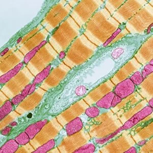
Trachea lining, SEM C013 / 7122
Trachea lining. Coloured scanning electron micrograph (SEM) of a section through the wall of a trachea (wind pipe). The trachea links the larynx to the lungs. The lining consists of mucus-secreting goblet cells (one seen at centre, red) and epithelial cells (vertical) that are covered in cilia (hair-like). Mucus traps debris, such as dust particles or bacteria, in the inhaled air, while the beating of the cilia moves the mucus and particles upwards out of the respiratory tract. This helps to keep the lungs and airways clear and prevent infection. Magnification: x3000 when printed at 10 centimetres wide
Science Photo Library features Science and Medical images including photos and illustrations
Media ID 9198651
© STEVE GSCHMEISSNER/SCIENCE PHOTO LIBRARY
Cilia Ciliated Cilium Colored Epithelial Epithelium Gland Glands Glandular Goblet Cell Lining Mucosal Mucosal Layer Mucous Membrane Protection Protective Respiratory Tract Secretion Secretory Subjects System Trachea Tract Wall Wind Pipe Cells Section Sectioned
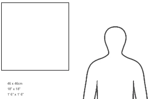
18"x18" (46x46cm) Pillow
18"x18" (46x46cm) Faux Suede Pillow with a plush soft feel. Your choice of image fills the front, with a stone colored faux suede back. Flat sewn concealed white zip.
Accessorise your space with decorative, soft pillows
Estimated Product Size is 45.7cm x 45.7cm (18" x 18")
These are individually made so all sizes are approximate
Artwork printed orientated as per the preview above, with landscape (horizontal) or portrait (vertical) orientation to match the source image.
EDITORS COMMENTS
This photo print, titled "Trachea lining, SEM C013 / 7122" offers a mesmerizing glimpse into the intricate world of our respiratory system. The image showcases a colored scanning electron micrograph (SEM) of a section through the wall of a trachea, commonly known as the windpipe. The lining of the trachea plays a vital role in protecting our lungs and airways from potential harm. It consists of mucus-secreting goblet cells, visible at the center in striking red hues, and vertically arranged epithelial cells covered in hair-like structures called cilia. The mucus produced by these goblet cells acts as an effective trap for debris present in the inhaled air, such as dust particles or bacteria. Simultaneously, the coordinated beating motion of the cilia propels this mucus along with trapped particles upwards and outwards from our respiratory tract. This crucial mechanism helps to keep our lungs clear and prevents infections from taking hold. With its magnification set at x3000 when printed at 10 centimeters wide, this photograph allows us to appreciate the remarkable complexity and beauty found within even microscopic aspects of human anatomy. Through images like this one captured by Steve Gschmeissner for Science Photo Library, we gain deeper insights into how our body's protective systems function on both macroscopic and microscopic levels.
MADE IN THE USA
Safe Shipping with 30 Day Money Back Guarantee
FREE PERSONALISATION*
We are proud to offer a range of customisation features including Personalised Captions, Color Filters and Picture Zoom Tools
SECURE PAYMENTS
We happily accept a wide range of payment options so you can pay for the things you need in the way that is most convenient for you
* Options may vary by product and licensing agreement. Zoomed Pictures can be adjusted in the Basket.