
Respiratory Tract Collection
The respiratory tract is a complex system within the human body that plays a vital role in our ability to breathe and maintain overall health


All Professionally Made to Order for Quick Shipping
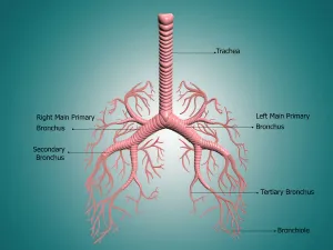
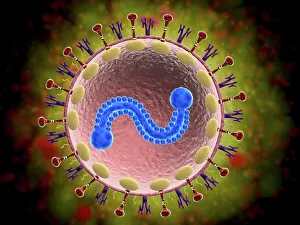
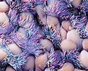
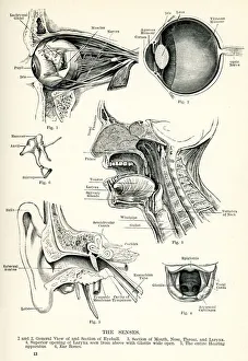
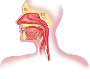
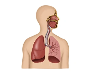
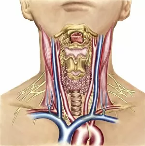
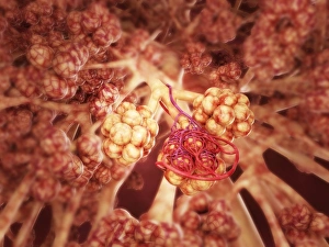
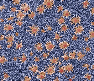
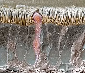
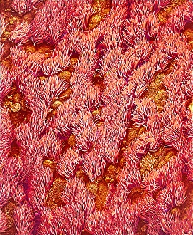
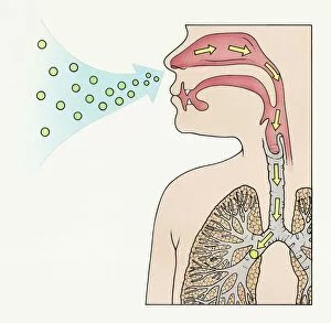
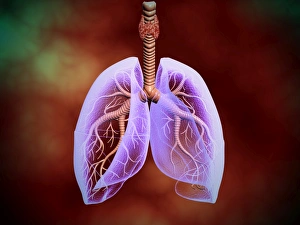
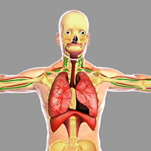
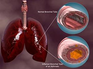
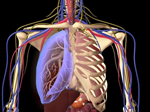
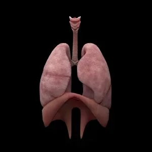
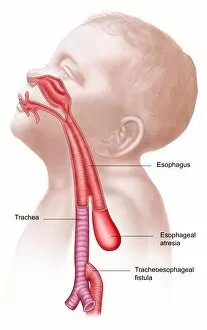
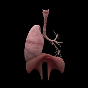
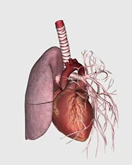
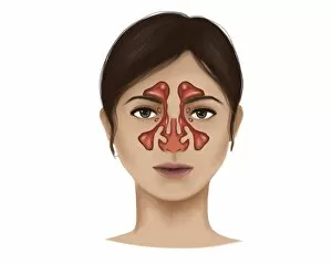
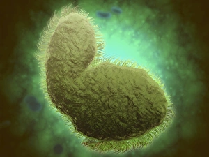
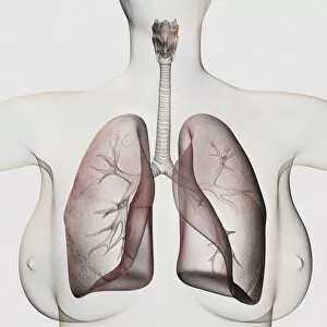
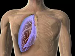
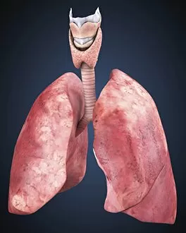
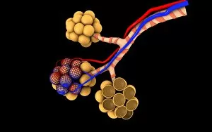
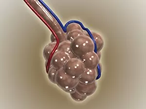
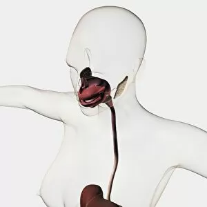
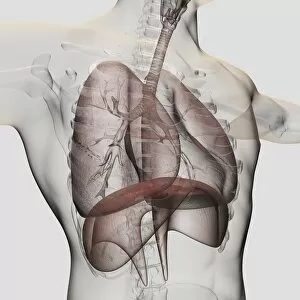
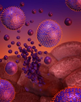
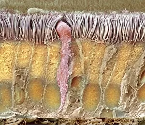
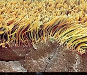
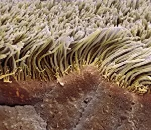
The respiratory tract is a complex system within the human body that plays a vital role in our ability to breathe and maintain overall health. From the anatomy of the bronchus and bronchial tubes to microscopic views of viruses like respiratory syncytial virus and adenovirus, this caption explores various aspects of this intricate network. Starting with an aerial view of a plane on a beach, we delve into the upper respiratory tract, depicted beautifully through artwork. Moving closer, we examine the nasal lining under a scanning electron microscope (SEM), revealing its delicate structure responsible for filtering air as it enters our bodies. Zooming further down, we encounter the trachea lining captured by SEM C013 / 7126. This image showcases the unique composition that lines our windpipe, protecting it from harmful particles while allowing smooth airflow. Continuing our journey through this incredible system, we explore both healthy and diseased alveoli in the lungs. These tiny sacs are crucial for gas exchange but can be affected by various conditions. As we conclude our exploration of the respiratory tract's anatomical wonders, let us remember how intertwined it is with other aspects of human physiology. It connects not only to organs directly involved in respiration but also to structures within our necks that support speech production and swallowing. In summary, these captivating images provide glimpses into different components of the respiratory tract – from macroscopic views like bronchial tubes to microscopic details such as viral infections or cellular linings. Understanding this intricate system helps us appreciate its importance in maintaining optimal lung function and overall well-being.