Photo Mug : Purkinje nerve cell, SEM


![]()
Home Decor from Science Photo Library
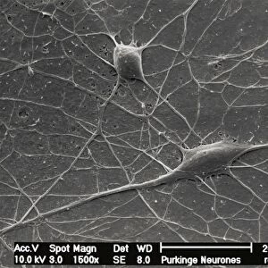
Purkinje nerve cell, SEM
Science Photo Library features Science and Medical images including photos and illustrations
Media ID 6421882
© DAVID MCCARTHY/SCIENCE PHOTO LIBRARY
Brown Cerebellum Connection Connections Dendrite Dendrites Granular Grey Matter Histology Junction Molecular Layer Nerve Cell Nervous Neuron Neurone Process Processes Purkinje System Tissue Brain Cells Neurology
Large Photo Mug (15 oz)
Brighten up your morning routine with our Media Storehouse Photo Mugs, featuring stunning scientific imagery. This mug showcases the intricate beauty of a Purkinje nerve cell, as captured in exquisite detail through Scanning Electron Microscopy from Science Photo Library. Each mug holds your favorite beverage while providing a daily dose of inspiration from the wonders of science. Perfect for scientists, students, or anyone who appreciates the beauty of the natural world.
Elevate your coffee or tea experience with our premium white ceramic mug. Its wide, comfortable handle makes drinking easy, and you can rely on it to be both microwave and dishwasher safe. Sold in single units, preview may show both sides of the same mug so you can see how the picture wraps around.
Elevate your coffee or tea experience with our premium white ceramic mug. Its wide, comfortable handle makes drinking easy, and you can rely on it to be both microwave and dishwasher safe. Sold in single units, preview may show both sides of the same mug so you can see how the picture wraps around.
These are individually made so all sizes are approximate
EDITORS COMMENTS
This print showcases the intricate beauty of a Purkinje nerve cell, captured using scanning electron microscopy (SEM). The image reveals the complex network of connections and processes that make up this vital component of our nervous system. In vibrant colors, we see the brown and white branches representing dendrites, which extend from the main body of the neuron known as the soma. These dendrites play a crucial role in receiving signals from other neurons and transmitting them to the soma. The molecular layer surrounding this Purkinje cell is depicted in shades of grey, highlighting its position within the cerebellum - an area responsible for motor control and coordination. This region is rich in granular cells, which can be seen forming connections with multiple dendrites. The detailed structure visible in this SEM image emphasizes both the complexity and elegance inherent in our neural architecture. It serves as a reminder of how essential these microscopic components are for maintaining a healthy functioning brain. As an invaluable tool for neurology research and histology studies, SEM allows us to explore these fascinating aspects of human anatomy on a cellular level. Science Photo Library has expertly captured this stunning representation that not only educates but also inspires awe at nature's design.
MADE IN THE USA
Safe Shipping with 30 Day Money Back Guarantee
FREE PERSONALISATION*
We are proud to offer a range of customisation features including Personalised Captions, Color Filters and Picture Zoom Tools
SECURE PAYMENTS
We happily accept a wide range of payment options so you can pay for the things you need in the way that is most convenient for you
* Options may vary by product and licensing agreement. Zoomed Pictures can be adjusted in the Cart.