Mouse Mat : Renal corpuscle of kidney

![]()
Home Decor From Science Photo Library
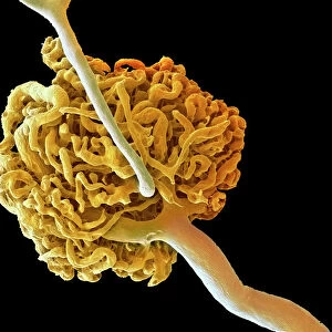
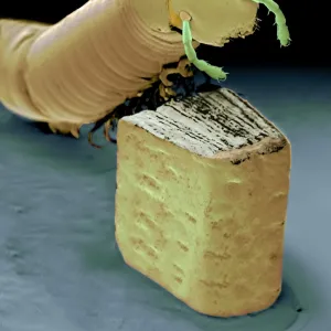
Renal corpuscle of kidney
Coloured scanning electron micrograph (SEM) of a renal corpuscle in the kidney. The renal corpuscle is formed by the glomerulus (red), the convoluted capillary at centre, which is surrounded by the Bowmans capsule, partly visible here as a white-brown menbrane at centre right. In the glomerulus blood is filtered. The filtrate is composed of waste and toxic products such as urea and chloride and nutrient substances. The filtrate is collected by the Bowmans capsule and then distributed to a network of tubules called nephrons two of which are visible coloured blue. Magnification: x330 at 6x7cm size
Science Photo Library features Science and Medical images including photos and illustrations
Media ID 6422712
© PROF. P. MOTTA/DEPT. OF ANATOMY/UNIVERSITY LA SAPIENZA , ROME/SCIENCE PHOTO LIBRARY
Blood Capillary Glomerulus Kidney Kidneys Magnified Image Microscopic Photos Nephron Subjects Urinary System False Coloured
Mouse Pad
Standard Size Mouse Pad 7.75" x 9..25". High density Neoprene w linen surface. Easy to clean, stain resistant finish. Rounded corners.
Archive quality photographic print in a durable wipe clean mouse mat with non slip backing. Works with all computer mice
Estimated Image Size (if not cropped) is 23.7cm x 19.6cm (9.3" x 7.7")
Estimated Product Size is 23.7cm x 20.2cm (9.3" x 8")
These are individually made so all sizes are approximate
Artwork printed orientated as per the preview above, with landscape (horizontal) orientation to match the source image.
EDITORS COMMENTS
This print showcases the intricate structure of a renal corpuscle in the kidney, captured using a coloured scanning electron microscope (SEM). The renal corpuscle consists of the glomerulus, depicted in vibrant red at the center, surrounded by Bowman's capsule. In this image, we can observe Bowman's capsule as a white-brown membrane towards the right. The glomerulus plays a crucial role in blood filtration within our bodies. As blood flows through it, waste and toxic products like urea and chloride are filtered out along with essential nutrients. This resulting mixture is known as filtrate. Bowman's capsule acts as an important collector for this filtrate before distributing it to nephrons – networks of tubules responsible for further processing and regulation within the urinary system. Two nephrons colored blue are visible in this magnified image. With a magnification level of x330 at 6x7cm size, this SEM photograph provides us with an extraordinary glimpse into the microscopic world hidden within our kidneys. It not only highlights the normal anatomy but also emphasizes its significance for maintaining overall human body health. Captured by Science Photo Library, renowned for their expertise in scientific imagery, this stunning print offers both educational value and aesthetic appeal to anyone interested in exploring subjects such as kidney function or microscopic photos.
MADE IN THE USA
Safe Shipping with 30 Day Money Back Guarantee
FREE PERSONALISATION*
We are proud to offer a range of customisation features including Personalised Captions, Color Filters and Picture Zoom Tools
SECURE PAYMENTS
We happily accept a wide range of payment options so you can pay for the things you need in the way that is most convenient for you
* Options may vary by product and licensing agreement. Zoomed Pictures can be adjusted in the Basket.