Metal Print : Pinned foot bones after surgery, X-ray

![]()
Metal Prints From Science Photo Library
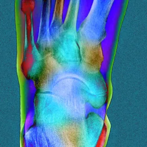
Pinned foot bones after surgery, X-ray
Pinned foot bones after surgery. Lateral (side view) X-ray showing pins (white) inserted into the bones of a patients foot. Pins have been inserted in to the heel bone (calcaneus), the tarsals and metatarsals, and the phalanges of the big toe. This type of surgery is done to correct deformities such as cavus foot, where the arch of the foot is too high. Pins and implants are also used in surgery to correct bunions and conditions such as hammer toe. For a foot like this being X-rayed during surgery, see image C014/7789. For an image showing insertion of a pin, see C014/7785
Science Photo Library features Science and Medical images including photos and illustrations
Media ID 9223495
© ANTONIA REEVE/SCIENCE PHOTO LIBRARY
Assessment Big Toe Bones Calcaneus Check Foot Heel Bone Implant Lateral Operation Orthopaedics Orthopedics Patient Pinned Pins Plate Plates Profile Radiography Surgery Surgical Tarsals Treatment X Ray Machine Xray
16"x20" (51x41cm) Metal Print
Discover the power of visual storytelling with Media Storehouse Metal Prints. This captivating image, "Pinned Foot Bones after Surgery" by Antonia Reeve from Science Photo Library, showcases a lateral X-ray revealing the intricacy of a patient's foot bones and the pins that hold them in place during the healing process. Each Metal Print is meticulously crafted with a vibrant, long-lasting finish that brings the detail and depth of this medical marvel to life. Add this unique piece to your home or office to inspire curiosity and spark conversation.
Made with durable metal and luxurious printing techniques, our metal photo prints go beyond traditional canvases, adding a cool, modern touch to your space. Wall mount on back. Eco-friendly 100% post-consumer recycled ChromaLuxe aluminum surface. The thickness of the print is 0.045". Featuring a Scratch-resistant surface and Rounded corners. Backing hangers are attached to the back of the print and float the print 1/2-inch off the wall when hung, the choice of hanger may vary depending on size and International orders will come with Float Mount hangers only. Finished with a brilliant white high gloss surface for unsurpassed detail and vibrance. Printed using Dye-Sublimation and for best care we recommend a non-ammonia glass cleaner, water, or isopropyl (rubbing) alcohol to prevent harming the print surface. We recommend using a clean, lint-free cloth to wipe off the print. The ultra-hard surface is scratch-resistant, waterproof and weatherproof. Avoid direct sunlight exposure.
Made with durable metal and luxurious printing techniques, metal prints bring images to life and add a modern touch to any space
Estimated Image Size (if not cropped) is 50.8cm x 40.6cm (20" x 16")
Estimated Product Size is 51.4cm x 41.2cm (20.2" x 16.2")
These are individually made so all sizes are approximate
Artwork printed orientated as per the preview above, with landscape (horizontal) orientation to match the source image.
EDITORS COMMENTS
This print showcases the remarkable aftermath of foot surgery, revealing a lateral (side view) X-ray of pinned foot bones. The image vividly displays white pins meticulously inserted into various sections of the patient's foot, including the heel bone (calcaneus), tarsals and metatarsals, as well as the phalanges of the big toe. This particular surgical procedure aims to rectify deformities such as cavus foot, characterized by an excessively high arch. The utilization of pins and implants in orthopedic surgeries extends beyond cavus foot correction; they also play a crucial role in addressing conditions like bunions and hammer toe. This photograph provides a glimpse into the intricate world of medical intervention for these ailments. The monochrome radiography highlights every detail with precision, offering healthcare professionals an invaluable assessment tool during post-surgical evaluations. Antonia Reeve's expert capture not only documents this specific case but also serves as a testament to advancements in orthopedics. It is important to note that this image does not represent any commercial use or affiliation with a company. Instead, it stands as a testament to human ingenuity and our unwavering commitment to improving lives through medicine and surgical innovation.
MADE IN THE USA
Safe Shipping with 30 Day Money Back Guarantee
FREE PERSONALISATION*
We are proud to offer a range of customisation features including Personalised Captions, Color Filters and Picture Zoom Tools
SECURE PAYMENTS
We happily accept a wide range of payment options so you can pay for the things you need in the way that is most convenient for you
* Options may vary by product and licensing agreement. Zoomed Pictures can be adjusted in the Basket.