
Implant Collection
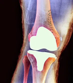
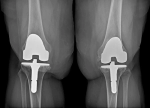
"Exploring the World of Implants: From Knee Joints to UFOs" Revolutionizing Mobility: Discover the wonders of knee joint prostheses and how they restore movement


All Professionally Made to Order for Quick Shipping
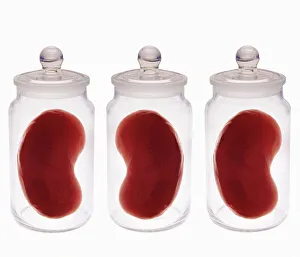
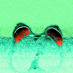
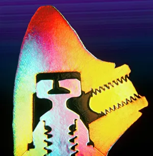
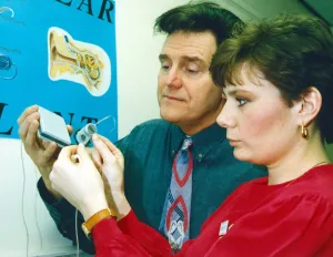
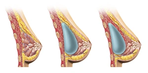
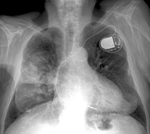
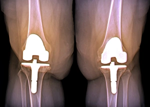
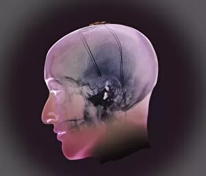
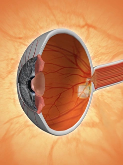
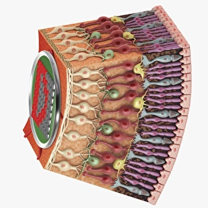
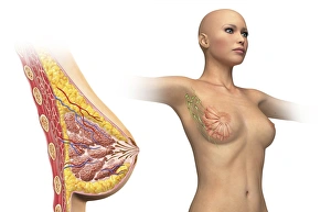
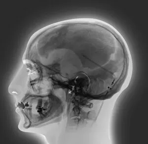
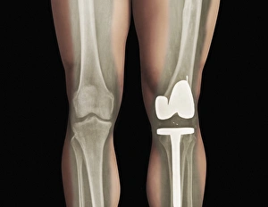
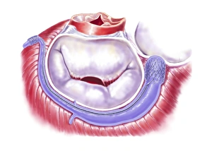
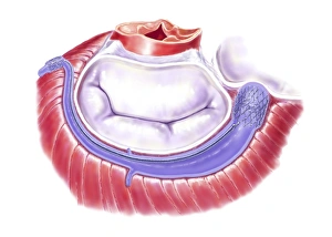
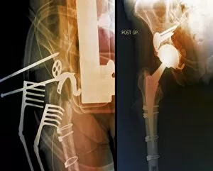
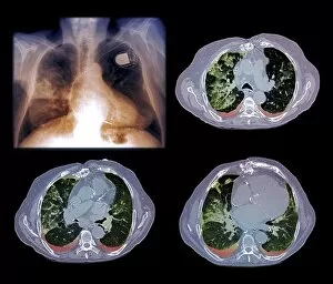
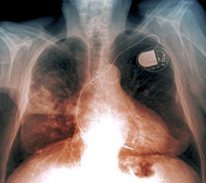
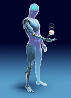
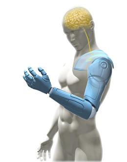
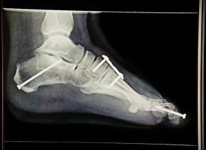
"Exploring the World of Implants: From Knee Joints to UFOs" Revolutionizing Mobility: Discover the wonders of knee joint prostheses and how they restore movement. Peek Inside: Unveiling the hidden secrets with X-ray technology, revealing the intricate world of implants. Spare Kidneys for All? Delving into the future possibilities of organ implants that could save lives. Out-of-This-World Encounters: Unraveling the mysterious connection between UFOs and alleged abductions involving implants. Safety Concerns: Investigating ruptured breast implants through advanced MRI scans, ensuring patient well-being. The Science Behind a Perfect Smile: Dive into a fascinating cross-section view of dental implant technology in action. Florian Sobocinski Hair Pieces Advert - Enhancing Confidence with Implant Solutions for Hair Loss AI meets Reality: Exploring body implants as part of augmented reality experiences powered by artificial intelligence in the cloud. A New Era Begins at North Riding Infirmary: Celebrating the opening of a unit dedicated to helping those with hearing impairments through innovative implant solutions. Beneath Beauty's Surface: Gaining insight into breast implant cross-sections, shedding light on cosmetic enhancements from within. Fighting Silent Battles: Detecting heart and lung diseases using X-ray imaging (X-ray C018/0498) – early diagnosis saves lives. Overcoming Obstacles Together: Analyzing X-rays (X-ray C016/6598) showcasing prosthetic knees' impact on obesity management and mobility improvement. 13. Prosthetic Knees & Weight Management Revealed. Witness how X-rays (X-ray C016/6596) highlight successful outcomes in battling obesity through knee replacements.