Jigsaw Puzzle : Glial cells, confocal light micrograph


![]()
Jigsaw Puzzles from Science Photo Library
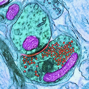
Glial cells, confocal light micrograph
Glial cells. Confocal light micrograph of glial cells from the cerebellum of the brain. Glial cells are nervous system cells that provide structural support and protection for neurons (nerve cells). Glial-fibrillary acidic protein, a type of intermediate filament (IF), part of the cells cytoskeleton, has been dyed yellow. The cytoskeleton is responsible for intracellular transport and cell structure and motility
Science Photo Library features Science and Medical images including photos and illustrations
Media ID 6299467
© THOMAS DEERINCK, NCMIR/SCIENCE PHOTO LIBRARY
Cell Biology Cerebellar Cerebellum Confocal Light Micrograph Cytology Cytoskeleton Fluorescence Fluorescent Gfap Glial Cell Glial Fibrillary Acidic Protein Intermediate Filament Neuroscience Support Cell Brain Cells Light Micrograph Light Microscope Protein
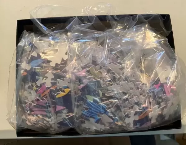
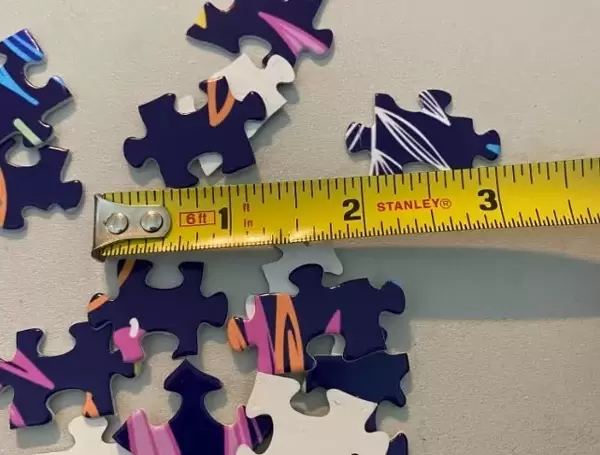
Jigsaw Puzzle (520 Pieces)
Discover the intricacies of the natural world with our Media Storehouse Jigsaw Puzzles. This captivating puzzle features a stunning confocal light micrograph image of Glial cells from the cerebellum of the brain, courtesy of Science Photo Library. Delve into the complex network of these essential nervous system cells, providing structural support and protection for neurons. Assemble this challenging jigsaw puzzle to enhance your knowledge and appreciation for the intricacies of the brain and neurobiology. Engage your mind, sharpen your focus, and unlock the secrets of the microscopic world - one piece at a time.
Made in the USA, 520-piece puzzles measure 16" x 20" (40.6 x 50.8 cm). Every puzzle is meticulously printed on glossy photo paper, which has a strong 1.33 mm thickness. Delivered in a black storage cardboard box, these puzzles are both stylish and practical. (Note: puzzles contain small parts and are not suitable for children under 3 years of age.)
Jigsaw Puzzles are an ideal gift for any occasion
Estimated Product Size is 40.5cm x 50.8cm (15.9" x 20")
These are individually made so all sizes are approximate
Artwork printed orientated as per the preview above, with landscape (horizontal) or portrait (vertical) orientation to match the source image.
EDITORS COMMENTS
This print showcases the intricate beauty of glial cells, captured through a confocal light micrograph. Found in the cerebellum of the brain, these glial cells play a crucial role in providing structural support and protection for neurons - the nerve cells that make up our nervous system. Highlighted in vibrant yellow is glial-fibrillary acidic protein (GFAP), which belongs to a group of intermediate filaments forming part of the cell's cytoskeleton. The cytoskeleton is responsible for various essential functions such as intracellular transport, maintaining cell structure, and facilitating motility. The image not only reveals the biological complexity within our brains but also emphasizes their healthy and normal state. Through fluorescence techniques, scientists have been able to visualize these vital support cells with exceptional clarity using a light microscope. This mesmerizing glimpse into cellular biology offers insights into neuroscience and cytology research. It serves as a reminder of how intricately interconnected our bodies are at even the smallest scale. Science Photo Library has once again provided us with an awe-inspiring visual representation of nature's wonders, allowing us to appreciate both its artistic value and scientific significance.
MADE IN THE USA
Safe Shipping with 30 Day Money Back Guarantee
FREE PERSONALISATION*
We are proud to offer a range of customisation features including Personalised Captions, Color Filters and Picture Zoom Tools
SECURE PAYMENTS
We happily accept a wide range of payment options so you can pay for the things you need in the way that is most convenient for you
* Options may vary by product and licensing agreement. Zoomed Pictures can be adjusted in the Cart.