Fine Art Print > Science Photo Library > Specialist Imaging
Fine Art Print : Glial stem cell culture, light micrograph

![]()
Fine Art Prints From Science Photo Library
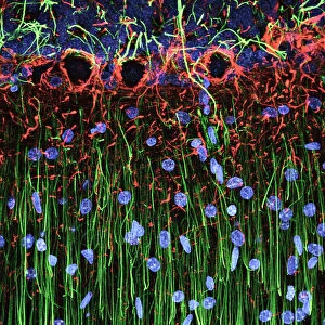
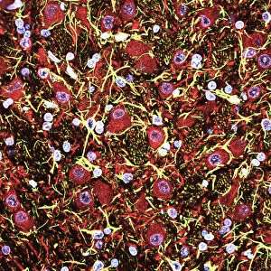
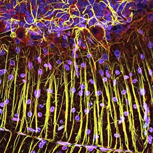
Glial stem cell culture, light micrograph
Glial stem cell culture. Fluorescent light micrograph of glial stem cells producing the protein NG2 (red) as they mature. These stem cells can differentiate into several types of glial cells, such as oligodendrocytes and astrocytes. More mature astrocyte progenitors are green, and nuclei are blue. Glial cells provide nerve cells with support and nutrition, form myelin (protein that coats the axons of the nerve cells) and participate in signal transmission in the nervous system. In the human brain, there are about 10 glia for each nerve cell
Science Photo Library features Science and Medical images including photos and illustrations
Media ID 6304709
© RICCARDO CASSIANI-INGONI/SCIENCE PHOTO LIBRARY
Astrocytes C Ulture Cell Biology Central Nervous System Confocal Cultured Cytology Dapi Developing Differentiating Differentiation Fluorescent Light Micrograph Glia Glial Cell Growing Histological Histology Multipotent Neuroglia Neuron Neurone Neurones Neurons Nuclei Nucleus Oligodendrocyte Progenitor Stem Cell Stem Cells Support Cell Cells Light Micrograph Light Microscope Neurological Neurology
20"x20" (+3" Border) Fine Art Print
Discover the intricacies of life at the cellular level with Media Storehouse's Fine Art Prints collection. This captivating image showcases a fluorescent light micrograph of glial stem cells in culture, as they mature and produce the protein NG2 (red). Witness the beauty and complexity of science with these exquisitely printed, museum-quality art pieces. Each print is meticulously crafted to bring out the vibrant colors and intricate details of this stunning science photograph from Science Photo Library, making it a perfect addition to any home or office space. Embrace the intersection of art and science with Media Storehouse's Fine Art Prints.
20x20 image printed on 26x26 Fine Art Rag Paper with 3" (76mm) white border. Our Fine Art Prints are printed on 300gsm 100% acid free, PH neutral paper with archival properties. This printing method is used by museums and art collections to exhibit photographs and art reproductions.
Our fine art prints are high-quality prints made using a paper called Photo Rag. This 100% cotton rag fibre paper is known for its exceptional image sharpness, rich colors, and high level of detail, making it a popular choice for professional photographers and artists. Photo rag paper is our clear recommendation for a fine art paper print. If you can afford to spend more on a higher quality paper, then Photo Rag is our clear recommendation for a fine art paper print.
Estimated Image Size (if not cropped) is 50.8cm x 50.8cm (20" x 20")
Estimated Product Size is 66cm x 66cm (26" x 26")
These are individually made so all sizes are approximate
Artwork printed orientated as per the preview above, with landscape (horizontal) or portrait (vertical) orientation to match the source image.
EDITORS COMMENTS
This print from Science Photo Library showcases the intricate world of glial stem cell culture. In this fluorescent light micrograph, we witness the remarkable process of maturation as glial stem cells produce the protein NG2, depicted in vibrant red hues. These versatile stem cells possess the extraordinary ability to differentiate into various types of glial cells, including oligodendrocytes and astrocytes. The more mature astrocyte progenitors are represented by a striking green coloration, while nuclei shimmer in brilliant shades of blue. Glial cells play a vital role in providing support and nourishment to nerve cells within the central nervous system (CNS). They also contribute to myelin formation – a crucial protein coating that envelops nerve cell axons – and actively participate in signal transmission throughout our complex neural network. It is fascinating to note that within the human brain, there exist approximately ten glia for every single nerve cell present. This image offers us an awe-inspiring glimpse into their world; a world where biological processes unfold with meticulous precision. As we delve deeper into this mesmerizing scene captured under a light microscope, we gain insight into cellular biology and cytology. The confocal imaging technique employed here allows us to observe differentiation occurring at its finest level – multipotent stem cells transforming into specialized support cells or progenitors. Science Photo Library has once again provided us with an exquisite visual representation of nature's wonders through this stunning photograph.
MADE IN THE USA
Safe Shipping with 30 Day Money Back Guarantee
FREE PERSONALISATION*
We are proud to offer a range of customisation features including Personalised Captions, Color Filters and Picture Zoom Tools
SECURE PAYMENTS
We happily accept a wide range of payment options so you can pay for the things you need in the way that is most convenient for you
* Options may vary by product and licensing agreement. Zoomed Pictures can be adjusted in the Basket.