
Premium Framed Print : Cells from a urine infection, SEM


![]()
Framed Photos From Science Photo Library
Cells from a urine infection, SEM
Cells from a urine infection. Coloured scanning electron micrograph (SEM) of crenated red blood cells, white blood cells and epithelial cells present in a urine sample taken from someone with a urinary tract infection (UTI). Magnification: x1650 when printed to centimetres wide
Science Photo Library features Science and Medical images including photos and illustrations
Media ID 6363457
© STEVE GSCHMEISSNER/SCIENCE PHOTO LIBRARY
Bladder Infection Colored Crenation Damage Damaged Dehydrated Epithelial Erythrocyte Erythrocytes Haematological Haematology Hematological Hematology Infected Infection Leucocyte Leucocytes Leukocyte Leukocytes Mycological Mycology Rbcs Red Blood Cell Sample Sediment Transitional Urinary Urinary Tract Infection Urological Urology White Blood Cell Abnormal Cells Crenate False Coloured Unhealthy
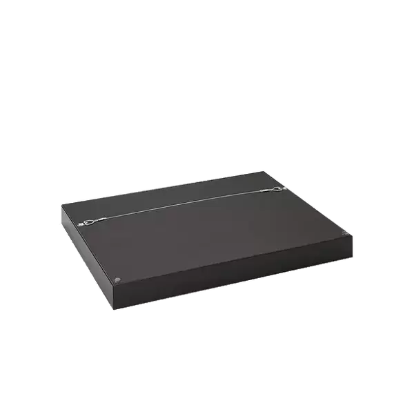
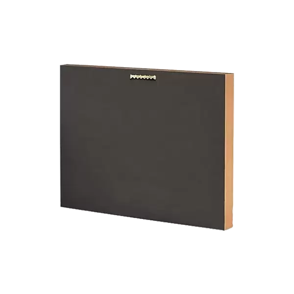
14"x16" Premium Frame
Contemporary style Premium Wooden Frame with 8"x10" Print. Complete with 2" White Mat and 1.25" thick MDF frame. Printed on 260 gsm premium paper. Glazed with shatter proof UV coated acrylic glass. Backing is paper covered backing with rubber bumpers. Supplied ready to hang with a pre-installed sawtooth/wire hanger. Care Instructions: Spot clean with a damp cloth. Securely packaged in a clear plastic bag and envelope in a reinforced cardboard shipper
FSC Real Wood Frame and Double Mounted with White Conservation Mountboard - Professionally Made and Ready to Hang
Estimated Image Size (if not cropped) is 25.4cm x 20.3cm (10" x 8")
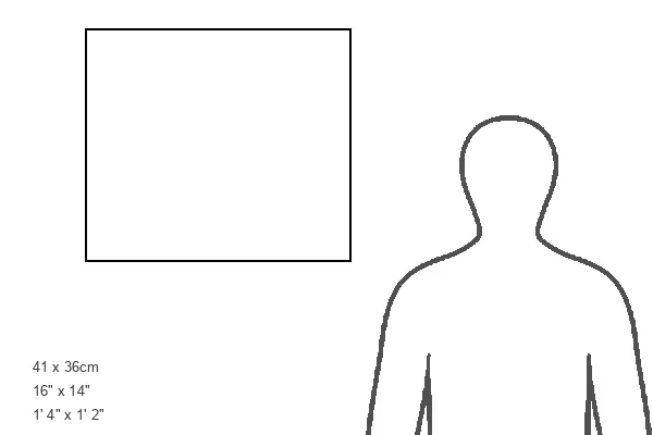
Estimated Product Size is 40.6cm x 35.6cm (16" x 14")
These are individually made so all sizes are approximate
Artwork printed orientated as per the preview above, with landscape (horizontal) orientation to match the source image.
EDITORS COMMENTS
This print from Science Photo Library showcases the intricate world of cells found in a urine infection. The image, captured using a scanning electron microscope (SEM), reveals an array of crenated red blood cells, white blood cells, and epithelial cells present in a urine sample taken from an individual suffering from a urinary tract infection (UTI). The vibrant colors used to highlight these microscopic entities add depth and visual appeal to this scientific marvel. With a magnification of x1650 when printed to centimeters wide, the level of detail is truly astonishing. Each cell tells its own story within this composition - damaged red blood cells hint at the toll the infection has taken on the body, while white blood cells stand as warriors fighting against invading pathogens. The presence of transitional epithelial cells further emphasizes the abnormality caused by this unhealthy condition. Beyond its aesthetic value, this image serves as an invaluable resource for researchers and medical professionals studying UTIs or related fields such as haematology, mycology, urology, and hematology. It offers insights into cellular structures that are otherwise invisible to our naked eye. Science Photo Library continues to provide exceptional visuals that bridge artistry with science. This particular photograph stands as a testament to their commitment in capturing moments that educate and inspire without commercial intent.
MADE IN THE USA
Safe Shipping with 30 Day Money Back Guarantee
FREE PERSONALISATION*
We are proud to offer a range of customisation features including Personalised Captions, Color Filters and Picture Zoom Tools
SECURE PAYMENTS
We happily accept a wide range of payment options so you can pay for the things you need in the way that is most convenient for you
* Options may vary by product and licensing agreement. Zoomed Pictures can be adjusted in the Basket.