Fraud Blocker Skip to main content

Leukocytes Collection

"Exploring the Dynamic World of Leukocytes: Unveiling their Intricate Structures and Functions" Plasma cells, captured under a TEM microscope

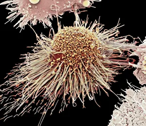
Background imageLeukocytes Collection: Activated macrophage, SEM C015 / 6375

Activated macrophage, SEM C015 / 6375
Activated macrophage. Coloured scanning electron micrograph (SEM) of an active macrophage white blood cell. Macrophages are cells of the bodys immune system that are found in the tissues rather than

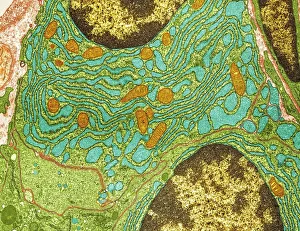
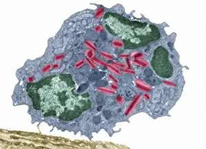
Background imageLeukocytes Collection: Plasma cells, TEM

Plasma cells, TEM
Plasma cells. Coloured transmission electron micrograph (TEM) of plasma cells. Plasma cells are mature B lymphocytes (white blood cells) that produce and secrete antibodies during an immune response

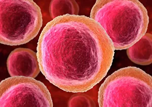
Background imageLeukocytes Collection: Lymphocyte white blood cells, artwork

Lymphocyte white blood cells, artwork
Lymphocyte white blood cells. Computer artwork of lymphocyte white blood cells, showing their nuclei (red, centre). Lymphocytes, like all white blood cells, are part of the immune system

Background imageLeukocytes Collection: White blood cells and platelets, SEM C016 / 3099

White blood cells and platelets, SEM C016 / 3099
White blood cells and platelets. Coloured scanning electron micrograph (SEM) of platelets (green) with a variety of different white blood cells (leucocytes, yellow)
Background imageLeukocytes Collection: White blood cells and platelets, SEM C016 / 3098

White blood cells and platelets, SEM C016 / 3098
White blood cells and platelets. Coloured scanning electron micrograph (SEM) of platelets (purple) with a variety of different white blood cells (leucocytes, blue)

Background imageLeukocytes Collection: Monocyte white blood cell, SEM C016 / 3089

Monocyte white blood cell, SEM C016 / 3089
Monocyte white blood cell. Scanning electron micrograph (SEM) of a monocyte white blood cell. Monocytes, like all white blood cells, are part of the human bodys immune system

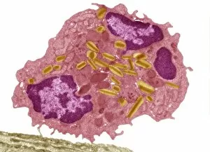
Background imageLeukocytes Collection: Eosinophil white blood cell, TEM C014 / 1438

Eosinophil white blood cell, TEM C014 / 1438
Eosinophil white blood cell. Transmission electron micrograph (TEM) of a section through an eosinophil. Eosinophils, like all white blood cells, are part of the bodys immune system

Background imageLeukocytes Collection: White blood cell and platelet, SEM F006 / 9929

White blood cell and platelet, SEM F006 / 9929
White blood cells. Coloured scanning electron micrograph (SEM) of white blood cells (leucocytes) and a platelet (thrombocyte, red). Magnification: x6, 600 when printed at 10 centimetres wide

Background imageLeukocytes Collection: White blood cell and platelet, SEM F006 / 9928

White blood cell and platelet, SEM F006 / 9928
White blood cells. Coloured scanning electron micrograph (SEM) of white blood cells (leucocytes) and a platelet (thrombocyte, orange). Magnification: x6, 600 when printed at 10 centimetres wide

Background imageLeukocytes Collection: White blood cell and platelet, SEM F006 / 9927

White blood cell and platelet, SEM F006 / 9927
White blood cells. Coloured scanning electron micrograph (SEM) of white blood cells (leucocytes) and a platelet (thrombocyte, green). Magnification: x6, 600 when printed at 10 centimetres wide

Background imageLeukocytes Collection: White blood cells, SEM F006 / 9924

White blood cells, SEM F006 / 9924
White blood cells. Coloured scanning electron micrograph (SEM) of white blood cells (leucocytes). Magnification: x2, 400 when printed at 10 centimetres wide

Background imageLeukocytes Collection: White blood cells, SEM F006 / 9923

White blood cells, SEM F006 / 9923
White blood cells. Coloured scanning electron micrograph (SEM) of white blood cells (leucocytes). Magnification: x2, 400 when printed at 10 centimetres wide

Background imageLeukocytes Collection: White blood cells, SEM F006 / 9926

White blood cells, SEM F006 / 9926
White blood cells. Coloured scanning electron micrograph (SEM) of white blood cells (leucocytes). Magnification: x2, 400 when printed at 10 centimetres wide

Background imageLeukocytes Collection: White blood cells, SEM F006 / 9925

White blood cells, SEM F006 / 9925
White blood cells. Coloured scanning electron micrograph (SEM) of white blood cells (leucocytes). Magnification: x2, 400 when printed at 10 centimetres wide

Background imageLeukocytes Collection: White blood cells, SEM F006 / 9922

White blood cells, SEM F006 / 9922
White blood cells. Coloured scanning electron micrograph (SEM) of white blood cells (leucocytes). Magnification: x2, 400 when printed at 10 centimetres wide

Background imageLeukocytes Collection: Eosinophil white blood cell, TEM C014 / 1437

Eosinophil white blood cell, TEM C014 / 1437
Eosinophil white blood cell. Transmission electron micrograph (TEM) of a section through an eosinophil. Eosinophils, like all white blood cells, are part of the bodys immune system

Background imageLeukocytes Collection: Eosinophil white blood cell, TEM C014 / 1439

Eosinophil white blood cell, TEM C014 / 1439
Eosinophil white blood cell. Transmission electron micrograph (TEM) of a section through an eosinophil. Eosinophils, like all white blood cells, are part of the bodys immune system

Background imageLeukocytes Collection: NK cells attacking cancer cells, artwork

NK cells attacking cancer cells, artwork
NK cells attacking cancer cells. Computer artwork of human Natural Killer (NK) cells (small, round) attacking cancer cells (large, round)

Background imageLeukocytes Collection: Antibodies and bacteria, artwork

Antibodies and bacteria, artwork
Antibodies and bacteria. Computer artwork showing white blood cells (large, round) and antibody, or immunoglobulin, molecules (red) surrounding bacteria (green)

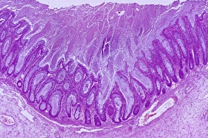
Background imageLeukocytes Collection: Colitis, light micrograph

Colitis, light micrograph
Colitis. Light micrograph of a section through colon (large intestine) tissue affected by bacterial infection, causing injury to the mucosal lining, which has resulted in inflammation (colitis)

Background imageLeukocytes Collection: Ischaemic bowel, light micrograph

Ischaemic bowel, light micrograph
Ischaemic bowel. Light micrograph of a section through a portion of intestine that has been damaged by an interruption of the blood supply, or ischaemia

Background imageLeukocytes Collection: Haematopoietic stem cells, artwork

Haematopoietic stem cells, artwork
Haematopoietic stem cells. Cutaway computer artwork showing white blood cells (leucocytes, white, round), red blood cells (erythrocytes, red) and haematopoietic stem cells (HSCs)

Background imageLeukocytes Collection: White blood cell antigen presentation C016 / 9058

White blood cell antigen presentation C016 / 9058
White blood cell antigen presentation. Coloured scanning electron micrograph (SEM) showing the interaction between a macrophage (yellow) and a T helper lymphocyte (Th cell, green)

Background imageLeukocytes Collection: Microglial white blood cell, SEM C016 / 9115

Microglial white blood cell, SEM C016 / 9115
Microglial white blood cell. Coloured scanning electron micrograph (SEM) of a microglial cell. Microglia are phagocytic macrophage white blood cells that are the main form of immune system in

Background imageLeukocytes Collection: Microglial white blood cell, SEM C016 / 9119

Microglial white blood cell, SEM C016 / 9119
Microglial white blood cell. Coloured scanning electron micrograph (SEM) of a microglial cell. Microglia are phagocytic macrophage white blood cells that are the main form of immune system in

Background imageLeukocytes Collection: Microglial white blood cell, SEM C016 / 9117

Microglial white blood cell, SEM C016 / 9117
Microglial white blood cell. Coloured scanning electron micrograph (SEM) of a microglial cell. Microglia are phagocytic macrophage white blood cells that are the main form of immune system in

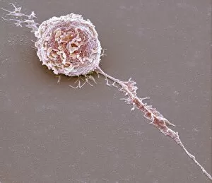
Background imageLeukocytes Collection: Lymphocyte white blood cell, SEM C016 / 9414

Lymphocyte white blood cell, SEM C016 / 9414
Lymphocyte white blood cell. Coloured scanning electron micrograph (SEM) of a lymphocyte white blood cell. Lymphocytes are involved in the immune systems defence mechanisms, lymph system

Background imageLeukocytes Collection: Microglial white blood cell, SEM C016 / 9109

Microglial white blood cell, SEM C016 / 9109
Microglial white blood cell. Coloured scanning electron micrograph (SEM) of a microglial cell. Microglia are phagocytic macrophage white blood cells that are the main form of immune system in

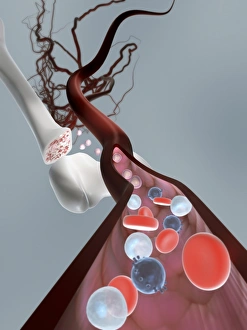
Background imageLeukocytes Collection: Human bone structure, artwork C016 / 7494

Human bone structure, artwork C016 / 7494
Human bone structure. Computer artwork showing all the different parts that make up a human bone, including the bone marrow (bottom left) where blood cells (round, white and red) are formed

Background imageLeukocytes Collection: Lymphocyte white blood cell, SEM C016 / 9415

Lymphocyte white blood cell, SEM C016 / 9415
Lymphocyte white blood cell. Coloured scanning electron micrograph (SEM) of a lymphocyte white blood cell. Lymphocytes are involved in the immune systems defence mechanisms, lymph system

Background imageLeukocytes Collection: Lymphocyte white blood cells, SEM C016 / 9140

Lymphocyte white blood cells, SEM C016 / 9140
Lymphocyte white blood cells. Coloured scanning electron micrograph (SEM) of lymphocyte white blood cells from peripheral blood

Background imageLeukocytes Collection: White blood cell antigen presentation C016 / 9057

White blood cell antigen presentation C016 / 9057
White blood cell antigen presentation. Coloured scanning electron micrograph (SEM) showing the interaction between a macrophage (red) and a T helper lymphocyte (Th cell, yellow)

Background imageLeukocytes Collection: Microglial white blood cell, SEM C016 / 9114

Microglial white blood cell, SEM C016 / 9114
Microglial white blood cell. Coloured scanning electron micrograph (SEM) of a microglial cell. Microglia are phagocytic macrophage white blood cells that are the main form of immune system in

Background imageLeukocytes Collection: Microglial white blood cells, SEM C016 / 9113

Microglial white blood cells, SEM C016 / 9113
Microglial white blood cells. Coloured scanning electron micrograph (SEM) of microglial cells. Microglia are phagocytic macrophage white blood cells that are the main form of immune system in

Background imageLeukocytes Collection: Microglial white blood cell, SEM C016 / 9118

Microglial white blood cell, SEM C016 / 9118
Microglial white blood cell. Coloured scanning electron micrograph (SEM) of a microglial cell. Microglia are phagocytic macrophage white blood cells that are the main form of immune system in

Background imageLeukocytes Collection: Microglial white blood cells, SEM C016 / 9111

Microglial white blood cells, SEM C016 / 9111
Microglial white blood cells. Coloured scanning electron micrograph (SEM) of microglial cells. Microglia are phagocytic macrophage white blood cells that are the main form of immune system in

Background imageLeukocytes Collection: Microglial white blood cells, SEM C016 / 9112

Microglial white blood cells, SEM C016 / 9112
Microglial white blood cells. Coloured scanning electron micrograph (SEM) of microglial cells. Microglia are phagocytic macrophage white blood cells that are the main form of immune system in

Background imageLeukocytes Collection: Microglial white blood cell, SEM C016 / 9116

Microglial white blood cell, SEM C016 / 9116
Microglial white blood cell. Coloured scanning electron micrograph (SEM) of a microglial cell. Microglia are phagocytic macrophage white blood cells that are the main form of immune system in

Background imageLeukocytes Collection: Lymphocyte white blood cells, SEM C016 / 9138

Lymphocyte white blood cells, SEM C016 / 9138
Lymphocyte white blood cells. Coloured scanning electron micrograph (SEM) of lymphocyte white blood cells from peripheral blood

Background imageLeukocytes Collection: Bone marrow blood cells, light micrograph

Bone marrow blood cells, light micrograph

Background imageLeukocytes Collection: Blood cells, light micrograph

Blood cells, light micrograph
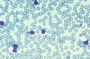
Blood cells. Light micrograph of red blood cells (erythrocytes, light blue) and white blood cells (leucocytes, nuclei stained purple)

Background imageLeukocytes Collection: Leukaemia cell, SEM

Leukaemia cell, SEM
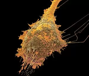
Leukaemia cell. Coloured scanning electron micrograph (SEM) of a leukaemic (cancerous) lymphocyte white blood cell (green), amongst normal red blood cells (erythrocytes, red)

Background imageLeukocytes Collection: Granulocyte blood cells, light micrograph

Granulocyte blood cells, light micrograph
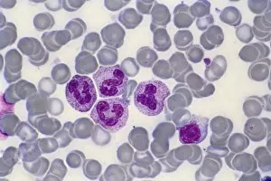
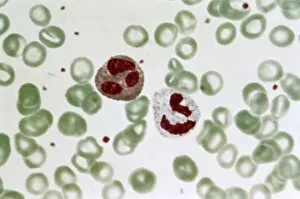
Granulocyte blood cells. Light micrograph of two types of granulocytes, a type of white blood cell (leucocyte). At left is a neutrophil, with an eosinophil at right

Background imageLeukocytes Collection: Antibodies and B cells, artwork C016 / 6255

Antibodies and B cells, artwork C016 / 6255
Antibodies and B cells. Computer artwork showing antibodies (green) neutralising cytokine activity in B cell (large, round) white blood cells

Background imageLeukocytes Collection: Antibodies and B cells, artwork C016 / 6254

Antibodies and B cells, artwork C016 / 6254
Antibodies and B cells. Computer artwork showing antibodies (green) neutralising cytokine activity in B cell (large, round) white blood cells

Background imageLeukocytes Collection: Normal and leukaemic blood, artwork C016 / 6261

Normal and leukaemic blood, artwork C016 / 6261
Normal and leukaemic blood. Computer artwork comparing the appearance of normal blood (left) and that affected by leukaemia (right)

Background imageLeukocytes Collection: Gastritis, light micrograph C016 / 0508

Gastritis, light micrograph C016 / 0508
Gastritis. Light micrograph of a section through an inflamed human stomach (gastritis), showing an abnormal accumulation of white blood cells (leucocytes)


All Professionally Made to Order for Quick Shipping

"Exploring the Dynamic World of Leukocytes: Unveiling their Intricate Structures and Functions" Plasma cells, captured under a TEM microscope, reveal their unique morphology and specialized role in producing antibodies to fight off infections. #MicroscopicMarvels Witness the remarkable transformation of an activated macrophage through SEM imaging (C015/6375). These powerful immune cells engulf and destroy harmful pathogens, safeguarding our bodies from harm. #MightyMacrophages Delve into the realm of lymphocyte white blood cells with captivating artwork that showcases their diversity and crucial role in orchestrating our body's immune response against invaders. #GuardiansOfImmunity Behold the intricate network formed by white blood cells and platelets as seen through SEM imaging (C016/3099). This symbiotic relationship ensures efficient clotting mechanisms while defending against foreign substances within our bloodstream. Dive deeper into the mesmerizing world of white blood cells and platelets with another stunning SEM image (C016/3098), revealing their incredible structures up close. #CellularWonders Explore the fascinating features of monocyte white blood cell under SEM microscopy (C016/3089) - these versatile warriors play a vital role in phagocytosis, clearing away debris, and initiating immune responses when needed. Picture No. 11675574 takes us on an extraordinary journey inside our bloodstream, showcasing various types of blood cells captured using high-resolution SEM technology – a testament to nature's beauty at its smallest scale. 8 & 9: Step into a microscopic wonderland where red and white blood cells dance together harmoniously as observed through breathtaking SEM images (#BloodCellsSEM). Marvel at this delicate balance essential for maintaining optimal health. 10: Eosinophil white blood cell steals the spotlight under TEM microscopy (C014/1438).