Fraud Blocker Skip to main content

Haematological Collection

"Exploring the Intricate World Marvels

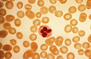
Background imageHaematological Collection: Dohle bodies in blood cell, micrograph

Dohle bodies in blood cell, micrograph
Dohle bodies in blood cell. Light micrograph of a neutrophil white blood cell (centre) with Dohle bodies. These are small inclusions within the cells cytoplasm

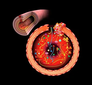
Background imageHaematological Collection: Blood coagulation cascade, artwork C016 / 9873

Blood coagulation cascade, artwork C016 / 9873
Blood coagulation cascade. Artwork of the biochemical cascade of blood chemicals and proteins during blood clotting (coagulation). The blood vessel and its layered wall is at upper left

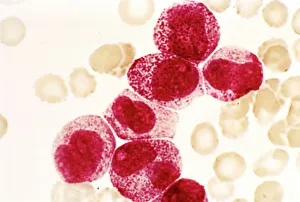
Background imageHaematological Collection: Acute promyelocytic leukaemia, micrograph

Acute promyelocytic leukaemia, micrograph
Acute promyelocytic leukaemia. Light micrograph of blood cells from bone marrow in a case of acute promyelocytic leukaemia. Leukaemia is a cancer where certain blood cells form in excess

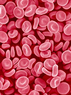
Background imageHaematological Collection: Red blood cells, SEM

Red blood cells, SEM
Red blood cells. Coloured scanning electron micrograph (SEM) of red blood cells (erythrocytes). Red blood cells are biconcave, disc-shaped cells that transport oxygen from the lungs to body cells

Background imageHaematological Collection: Lymphocyte white blood cells, artwork

Lymphocyte white blood cells, artwork
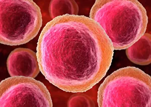
Lymphocyte white blood cells. Computer artwork of lymphocyte white blood cells, showing their nuclei (red, centre). Lymphocytes, like all white blood cells, are part of the immune system

Background imageHaematological Collection: Haematopoietic stem cells, SEM C013 / 5009

Haematopoietic stem cells, SEM C013 / 5009
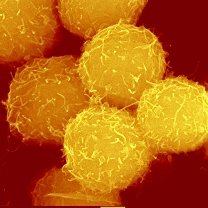
Haematopoietic stem cells, coloured scanning electron micrograph (SEM). Stem cells can differentiate into any other cell type

Background imageHaematological Collection: Myeloblast blood cell, light micrograph

Myeloblast blood cell, light micrograph
Myeloblast blood cell. Light micrograph of blood cells, including a myeloblast, a precursor for a type of white blood cell (leucocyte). Myeloblasts differentiate into granulocytes

Background imageHaematological Collection: Promyelocyte blood cell, light micrograph

Promyelocyte blood cell, light micrograph. This blood cell (centre) is a precursor for a type of white blood cell called a granulocyte, formed by granulopoiesis in the bone marrow

Background imageHaematological Collection: Chronic lymphocytic leukaemia, micrograph

Chronic lymphocytic leukaemia, micrograph
Chronic lymphocytic leukaemia. Light micrograph of blood cells sampled from a lymphatic ganglion in a case of chronic lymphocytic leukaemia, also called chronic lymphoid leukaemia (CLL)

Background imageHaematological Collection: Macrophage and lymphocytes, TEM

Macrophage and lymphocytes, TEM
Macrophage and lymphocytes, transmission electron micrograph (TEM). Macrophage surrounded by lymphocytes in a lymph node. This view includes many intercellular contacts

Background imageHaematological Collection: White blood cells and platelets, SEM C016 / 3099

White blood cells and platelets, SEM C016 / 3099
White blood cells and platelets. Coloured scanning electron micrograph (SEM) of platelets (green) with a variety of different white blood cells (leucocytes, yellow)

Background imageHaematological Collection: White blood cells and platelets, SEM C016 / 3098

White blood cells and platelets, SEM C016 / 3098
White blood cells and platelets. Coloured scanning electron micrograph (SEM) of platelets (purple) with a variety of different white blood cells (leucocytes, blue)

Background imageHaematological Collection: Monocyte white blood cell, SEM C016 / 3089

Monocyte white blood cell, SEM C016 / 3089
Monocyte white blood cell. Scanning electron micrograph (SEM) of a monocyte white blood cell. Monocytes, like all white blood cells, are part of the human bodys immune system

Background imageHaematological Collection: Red blood cells, light micrograph C016 / 3035

Red blood cells, light micrograph C016 / 3035
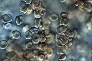
Red blood cells. Differential interference contrast (DIC) micrograph of red blood cells (erythrocytes). Red blood cells are biconcave, giving them a large surface area for gas exchange

Background imageHaematological Collection: Thrombosed blood vessel, artwork C013 / 4649

Thrombosed blood vessel, artwork C013 / 4649
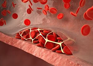
Thrombosed blood vessel. Computer artwork of a blood clot (thrombus) formed on the internal wall of a blood vessel

Background imageHaematological Collection: Blood samples C018 / 1034

Blood samples C018 / 1034
Blood samples

Background imageHaematological Collection: Blood smears F008 / 2107

Blood smears F008 / 2107
Blood smears on microscope slides

Background imageHaematological Collection: Haematology lab F007 / 0384

Haematology lab F007 / 0384
MODEL RELEASED. Haematology lab. Laboratory assistant using a multipipette to transfer blood samples

Background imageHaematological Collection: Haematology lab F007 / 0293

Haematology lab F007 / 0293
MODEL RELEASED. Haematology lab. Laboratory assistant using a pipette to transfer blood samples

Background imageHaematological Collection: Haematology lab F007 / 0288

Haematology lab F007 / 0288
MODEL RELEASED. Haematology lab. Laboratory assistant examining blood samples in a multiwell tray

Background imageHaematological Collection: Haematology lab F007 / 0287

Haematology lab F007 / 0287
MODEL RELEASED. Haematology lab. Laboratory assistant using a pipette to transfer blood samples

Background imageHaematological Collection: Haematology lab F007 / 0286

Haematology lab F007 / 0286
MODEL RELEASED. Haematology lab. Laboratory assistant pipetting a blood sample onto a slide

Background imageHaematological Collection: Haematology lab F007 / 0284

Haematology lab F007 / 0284
MODEL RELEASED. Haematology lab. Laboratory assistant using a multipipette to transfer blood samples

Background imageHaematological Collection: Haematology lab F007 / 0285

Haematology lab F007 / 0285
MODEL RELEASED. Haematology lab. Laboratory assistant using a pipette to transfer blood samples

Background imageHaematological Collection: Haemoglobin, molecular model F006 / 9604

Haemoglobin, molecular model F006 / 9604
Haemoglobin, molecular model. This is deoxyhaemoglobin, the molecule in its non-oxygen bound state. Haemoglobin transports oxygen around the body in red blood cells

Background imageHaematological Collection: Haemoglobin S, molecular model F006 / 9601

Haemoglobin S, molecular model F006 / 9601
Haemoglobin S. Molecular model of the mutant form of haemoglobin (haemoglobin S) that causes sickle cell anaemia. This is deoxyhaemoglobin S, the molecule in its non-oxygen bound state

Background imageHaematological Collection: Haemoglobin, molecular model F006 / 9580

Haemoglobin, molecular model F006 / 9580
Haemoglobin, molecular model. This is deoxyhaemoglobin, the molecule in its non-oxygen bound state. Haemoglobin transports oxygen around the body in red blood cells

Background imageHaematological Collection: Thrombin complexed with fibrinogen F006 / 9561

Thrombin complexed with fibrinogen F006 / 9561
Thrombin complexed with fibrinogen, molecular model. The thrombin molecules (beige, right and left) are bound to the central part of the fibrinogen molecule (down centre)

Background imageHaematological Collection: Haemoglobin molecule F006 / 9356

Haemoglobin molecule F006 / 9356
Haemoglobin, molecular model. Haemoglobin is a metalloprotein that transports oxygen around the body in red blood cells. Each molecule consists of iron-containing haem groups (sticks)

Background imageHaematological Collection: Haemoglobin molecule F006 / 9350

Haemoglobin molecule F006 / 9350
Haemoglobin, molecular model. Haemoglobin is a metalloprotein that transports oxygen around the body in red blood cells. Each molecule consists of iron-containing haem groups (sticks)

Background imageHaematological Collection: Blood sample F006 / 9054

Blood sample F006 / 9054
Blood sample and syringe

Background imageHaematological Collection: Blood sample F006 / 9055

Blood sample F006 / 9055
Blood sample

Background imageHaematological Collection: Haematopoietic stem cells, artwork

Haematopoietic stem cells, artwork
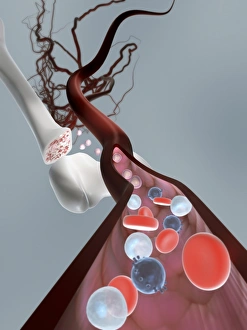
Haematopoietic stem cells. Cutaway computer artwork showing white blood cells (leucocytes, white, round), red blood cells (erythrocytes, red) and haematopoietic stem cells (HSCs)

Background imageHaematological Collection: Thickened skull, MRI scan

Thickened skull, MRI scan
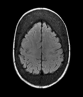
Thickened skull. Magnetic resonance imaging (MRI) scan of an axial section through the head of a 22-year-old patient, showing thickening of the trabecular (spongy) bone of the skull

Background imageHaematological Collection: Atherosclerosis, 3D CT scan

Atherosclerosis, 3D CT scan
Atherosclerosis. 3D computed tomography (CT) angiogram of the main blood vessels of the lower body a 58-year-old man, showing numerous atheromatous plaques (light, centre) on the vessel walls

Background imageHaematological Collection: White blood cell antigen presentation C016 / 9058

White blood cell antigen presentation C016 / 9058
White blood cell antigen presentation. Coloured scanning electron micrograph (SEM) showing the interaction between a macrophage (yellow) and a T helper lymphocyte (Th cell, green)

Background imageHaematological Collection: Microglial white blood cell, SEM C016 / 9115

Microglial white blood cell, SEM C016 / 9115
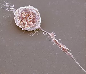
Microglial white blood cell. Coloured scanning electron micrograph (SEM) of a microglial cell. Microglia are phagocytic macrophage white blood cells that are the main form of immune system in

Background imageHaematological Collection: Light microscopy C016 / 9700

Light microscopy C016 / 9700
Light microscopy. Light microscope being used to magnify a blood sample

Background imageHaematological Collection: Blood clot, SEM C016 / 9088

Blood clot, SEM C016 / 9088
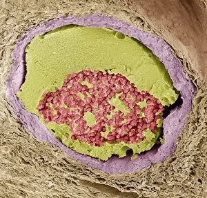
Blood clot. Coloured scanning electron micrograph (SEM) of a section through a blood clot (thrombus) in an arteriole (small blood vessel), showing red blood cells (red) in a fibrin mesh (green)

Background imageHaematological Collection: Microglial white blood cell, SEM C016 / 9119

Microglial white blood cell, SEM C016 / 9119
Microglial white blood cell. Coloured scanning electron micrograph (SEM) of a microglial cell. Microglia are phagocytic macrophage white blood cells that are the main form of immune system in

Background imageHaematological Collection: Microglial white blood cell, SEM C016 / 9117

Microglial white blood cell, SEM C016 / 9117
Microglial white blood cell. Coloured scanning electron micrograph (SEM) of a microglial cell. Microglia are phagocytic macrophage white blood cells that are the main form of immune system in

Background imageHaematological Collection: Platelet, SEM C016 / 9060

Platelet, SEM C016 / 9060
Platelet, coloured scanning electron micrograph (SEM). Platelets, or thrombocytes, are fragments of white blood cells. When blood clots

Background imageHaematological Collection: Lymphocyte white blood cell, SEM C016 / 9414

Lymphocyte white blood cell, SEM C016 / 9414
Lymphocyte white blood cell. Coloured scanning electron micrograph (SEM) of a lymphocyte white blood cell. Lymphocytes are involved in the immune systems defence mechanisms, lymph system

Background imageHaematological Collection: Microglial white blood cell, SEM C016 / 9109

Microglial white blood cell, SEM C016 / 9109
Microglial white blood cell. Coloured scanning electron micrograph (SEM) of a microglial cell. Microglia are phagocytic macrophage white blood cells that are the main form of immune system in

Background imageHaematological Collection: Light microscopy C016 / 9703

Light microscopy C016 / 9703
Light microscopy. Light microscope being used to magnify a blood sample

Background imageHaematological Collection: Crenated red blood cells, SEM C016 / 9029

Crenated red blood cells, SEM C016 / 9029
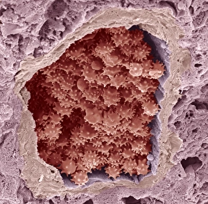
Crenated red blood cells. Coloured scanning electron micrograph (SEM) of a section through an arteriole (small blood vessel), showing crenated red blood cells inside

Background imageHaematological Collection: Human bone structure, artwork C016 / 7494

Human bone structure, artwork C016 / 7494
Human bone structure. Computer artwork showing all the different parts that make up a human bone, including the bone marrow (bottom left) where blood cells (round, white and red) are formed

Background imageHaematological Collection: Blood clot, SEM C016 / 9089

Blood clot, SEM C016 / 9089
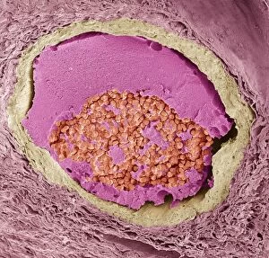
Blood clot. Coloured scanning electron micrograph (SEM) of a section through a blood clot (thrombus) in an arteriole (small blood vessel), showing red blood cells (red) in a fibrin mesh (bright pink)


All Professionally Made to Order for Quick Shipping

"Exploring the Intricate World Marvels: From Blood Coagulation to Leukaemia" Delve into the mesmerizing realm of haematology with a captivating artwork showcasing the intricate blood coagulation cascade (Artwork C016 / 9873). Witness the enigmatic Dohle bodies within a blood cell through a powerful micrograph, unraveling their significance in haematological diagnostics. Behold the stunning beauty of red blood cells captured by a scanning electron microscope (SEM), revealing their unique structure and function. Uncover the haunting reality of Acute Promyelocytic Leukaemia through an evocative micrograph, shedding light on this devastating blood disorder. Immerse yourself in an artistic portrayal of lymphocyte white blood cells, celebrating their vital role in our immune system's defense against pathogens (Artwork). Get up close and personal with a striking SEM image capturing the intricate details of a blood clot formation, highlighting its crucial role in wound healing and preventing excessive bleeding. Explore the fascinating world of haematopoietic stem cells through a remarkable SEM image (C013 / 5009), marveling at their ability to give rise to all types of mature blood cells. Journey into the microscopic realm as you witness myeloblasts and promyelocytes under light microscopy, providing insights into early stages of white blood cell development. Peer into an extraordinary TEM image depicting macrophages interacting with lymphocytes, offering glimpses into how these immune cells collaborate for optimal defense against infections. Experience awe-inspiring SEM images capturing white blood cells alongside platelets (C016 / 3099 & C016 / 3098), illustrating their collective effort in maintaining our body's delicate balance between immunity and clotting mechanisms.