Photographic Print : Moss spore capsule, light micrograph

![]()
Photo Prints From Science Photo Library
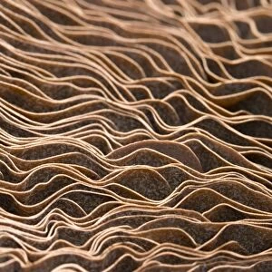
Moss spore capsule, light micrograph
Moss spore capsule, polarised light micrograph. Longitudinal section through a spore capsule from a fire moss (Funaria hygrometrica). The capsules cover is dark green at its base (apophysis), where photosynthetic tissue makes food for the developing spores. The inner capsule (upper centre) is where the spores form. The inner capsule has a lid (operculum, pink), with edges (annulus, bright green) that break down to allow the lid to fall off, releasing the spores. Magnification: x45 when printed at 10 centimetres high
Science Photo Library features Science and Medical images including photos and illustrations
Media ID 6308029
© DR KEITH WHEELER/SCIENCE PHOTO LIBRARY
Bryophyte Bryophytes Bryophytic Cellular Internal Structure Longitudinal Moss Mosses Operculum Orange Orange Background Part Parts Plant Anatomy Polarised Polarized Re Production Reproductive Reproductive Part Reproductive Parts Sporangium Spore Spore Capsule Spores Structural Tissue Annulus Cells Light Micrograph Light Microscope Section Sectioned
12"x8" Photo Print
Discover the intricacy of nature with Media Storehouse's Photographic Prints. This captivating image showcases a Moss Spore Capsule, captured in stunning detail through a polarized light micrograph by Science Photo Library. A longitudinal section of the Fire Moss (Funaria hygrometrica) spore capsule reveals a mesmerizing labyrinth of intricately patterned cells. Bring this microscopic marvel into your home or office as a conversation starter and a reminder of the beauty hidden in the smallest corners of our world.
Photo prints are produced on Kodak professional photo paper resulting in timeless and breath-taking prints which are also ideal for framing. The colors produced are rich and vivid, with accurate blacks and pristine whites, resulting in prints that are truly timeless and magnificent. Whether you're looking to display your prints in your home, office, or gallery, our range of photographic prints are sure to impress. Dimensions refers to the size of the paper in inches.
Our Photo Prints are in a large range of sizes and are printed on Archival Quality Paper for excellent colour reproduction and longevity. They are ideal for framing (our Framed Prints use these) at a reasonable cost. Alternatives include cheaper Poster Prints and higher quality Fine Art Paper, the choice of which is largely dependant on your budget.
Estimated Image Size (if not cropped) is 16.4cm x 30.4cm (6.5" x 12")
Estimated Product Size is 20.3cm x 30.5cm (8" x 12")
These are individually made so all sizes are approximate
Artwork printed orientated as per the preview above, with portrait (vertical) orientation to match the source image.
EDITORS COMMENTS
This print showcases the intricate beauty of a moss spore capsule, captured under polarized light microscopy. The image provides a longitudinal section view of a spore capsule from the fire moss species known as Funaria hygrometrica. At its base, the dark green cover called apophysis houses photosynthetic tissue responsible for nourishing the developing spores. In the upper center, we can observe the inner capsule where these spores take shape. The pink lid, scientifically referred to as operculum, rests atop the inner capsule and is surrounded by bright green edges called annulus. As part of their reproductive process, these annulus edges gradually break down to allow the lid to detach and release an abundance of tiny spores into their surroundings. Printed at 10 centimeters high with a magnification factor of x45, this image offers us an up-close look at nature's microscopic wonders. It unveils not only the internal structure but also highlights various botanical elements such as cells and tissues that play crucial roles in moss reproduction. With its vibrant orange background contrasting against delicate shades of green and pink, this photograph serves as a testament to both scientific exploration and artistic appreciation for plant anatomy.
MADE IN THE USA
Safe Shipping with 30 Day Money Back Guarantee
FREE PERSONALISATION*
We are proud to offer a range of customisation features including Personalised Captions, Color Filters and Picture Zoom Tools
SECURE PAYMENTS
We happily accept a wide range of payment options so you can pay for the things you need in the way that is most convenient for you
* Options may vary by product and licensing agreement. Zoomed Pictures can be adjusted in the Basket.