Fraud Blocker Skip to main content

Re Production Collection

Exploring the wonders of reproduction through time and nature

Background imageRe Production Collection: Catalan Atlas, 14th century

Catalan Atlas, 14th century. The 6-page Catalan Atlas (1375) was produced on vellum by the Jewish cartographer Abraham Cresques

Background imageRe Production Collection: Da Vincis crossbow

Da Vincis crossbow. Late 19th Century reproduction of a page from a notebook written by Leonardo da Vinci (1452-1519), showing his design for a giant crossbow. Published circa 1894-1904

Background imageRe Production Collection: Tyrannosaurus rex dinosaurs mating

Tyrannosaurus rex dinosaurs mating. Artwork of male (right) and female (left) Tyrannosaurus rex dinosaurs mating. Some theories say that this dinosaur had feathers and fur, as seen here

Background imageRe Production Collection: Uterus lining during menstruation, SEM

Uterus lining during menstruation, SEM
Uterus during menstruation. Coloured scanning electron micrograph (SEM) of the lining of the uterus being shed during menstruation

Background imageRe Production Collection: Illustration of the human placenta

Illustration of the human placenta. The placenta is an organ that develops in the female uterus during pregnancy. Through it, and an umbilical cord (at lower right)

Background imageRe Production Collection: English oak (Quercus robur) acorns

English oak (Quercus robur) acorns. Acorns forming on an English oak tree. This tree is also known as the pedunculate oak. Acorns are the fruit of the oak tree

Background imageRe Production Collection: Geranium anther, SEM

Geranium anther, SEM
Geranium anther. Coloured scanning electron micrograph (SEM) of pollen (pink) on the anther of a geranium flower (Geranium sp.)

Background imageRe Production Collection: Dahlia flower pollen, SEM

Dahlia flower pollen, SEM
Dahlia flower pollen. Coloured scanning electron micrograph (SEM) of a pollen grain (orange) from a Dahlia cultorum flower. A pollen grain contains a male sex cell from a flowering plant

Background imageRe Production Collection: Maple seed flight path

Maple seed flight path. Multi-flash photo composition of a maple seed (Acer sp.) spinning as it falls. Maple seeds are attached to the tree in pairs

Background imageRe Production Collection: STEVENSON, Robert Louis (1850-1894). Reproduction

STEVENSON, Robert Louis (1850-1894). Reproduction of a picture of Stevenson taken in 1885

Background imageRe Production Collection: CRESQUES, Jafuda (1350-1410); CRESQUES, Abraham

CRESQUES, Jafuda (1350-1410); CRESQUES, Abraham ( -1381). Catalan Atlas. 1375. Third and Fourth Leaves. Map of the world known in the 14th c. (Finisterre and the Mediterranean)

Background imageRe Production Collection: Sea cucumber

Sea cucumber (Thelenota sp.) spawning. Microscopic sex cells (gametes) are being released to allow fertilisation (the fusing of a male and a female sex cell)

Background imageRe Production Collection: Pollen grains, SEM

Pollen grains, SEM
Pollen grains. Coloured scanning electron micrographs (SEM) of pollen grains from a variety of plants

Background imageRe Production Collection: Cloudberry (Rubus chamaemorus)

Cloudberry (Rubus chamaemorus)
Cloudberry fruit (Rubus chamaemorus)

Background imageRe Production Collection: Nelsons last signal at Trafalgar, 21 October 1805

Nelsons last signal at Trafalgar, 21 October 1805
At the Battle of Trafalgar on 21 October 1805, Nelson gives his famous last signal England expects that every man will do his duty

Background imageRe Production Collection: Herpes virus replicating

Herpes virus replicating, computer artwork.Viruses are only able to replicate in a host cell.The glycoprotein spikes in the virus envelope(green)

Background imageRe Production Collection: Easter cactus stigma, SEM

Easter cactus stigma, SEM
Easter cactus stigma. Coloured scanning electron micrograph (SEM) of the stigma (pink) and style (green) of an Easter cactus flower (Rhipsalidopsis gaertneri)

Background imageRe Production Collection: Candida fungus, SEM

Candida fungus, SEM
Candida fungus. Coloured scanning electron micrograph (SEM) of budding threads (hyphae) of a Candida fungus. The budding areas are where asexual reproduction is producing new fungus cells

Background imageRe Production Collection: Lactating breast tissue, light micrograph

Lactating breast tissue, light micrograph
Lactating breast tissue. Light micrograph of a section through healthy breast (mammary gland) tissue during lactation. Glandular tissue in the breast has proliferated to produce spaces (alveoli)

Background imageRe Production Collection: Pregnant woman

Pregnant woman holding her aching back

Background imageRe Production Collection: Honeybee leg, SEM

Honeybee leg, SEM
Honey bee leg. Coloured scanning electron micrograph (SEM) of the leg of a honeybee (Apis mellifera) covered in pollen grains (round). The dense coating of hairs has trapped the pollen

Background imageRe Production Collection: Mushroom gills, SEM

Mushroom gills, SEM
Mushroom gills. Coloured scanning electron micrograph (SEM) of the gills of an unidentified gilled mushroom. The mushroom is the fruiting body of a fungus

Background imageRe Production Collection: Dividing yeast cells, SEM

Dividing yeast cells, SEM
Dividing yeast cells. Coloured scanning electron micrograph (SEM) of Schizosaccharomyces pombe yeast cells dividing. S. pombe is a single-celled fungus that is studied widely as a model organism for

Background imageRe Production Collection: Dividing yeast cells, SEM

Dividing yeast cells, SEM
Dividing yeast cells. Coloured scanning electron micrograph (SEM) of Schizosaccharomyces pombe yeast cells dividing. S. pombe is a single-celled fungus that is studied widely as a model organism for

Background imageRe Production Collection: Tea flower stamens, SEM

Tea flower stamens, SEM
Tea flower stamens. Coloured scanning electron micrograph (SEM) of the stamens of a flower of the tea plant (Camellia sp.). Stamens are the male reproductive structures of a flower

Background imageRe Production Collection: Delphinium flowers

Delphinium flowers
Delphinium (Delphinium Blue Indulgence ) flowers

Background imageRe Production Collection: Green turtles mating

Green turtles mating. The green turtle (Chelonia mydas) is critically under threat of extinction and is a protected species. Photographed off Sipadan, Sabah, Malaysia

Background imageRe Production Collection: Portrait of 1st Viscount Horatio Nelson, 1785-1805

Portrait of 1st Viscount Horatio Nelson, 1785-1805
This is a photographic reproduction of a painting of the 1st Viscount Horatio Nelson who lived from 1758 to 1805

Background imageRe Production Collection: Souvenirs for sale, Rome, Lazio, Italy, Europe

Souvenirs for sale, Rome, Lazio, Italy, Europe

Background imageRe Production Collection: Geranium pollen, SEM

Geranium pollen, SEM
Geranium pollen. Coloured scanning electron micrograph (SEM) of pollen (pink) on the stamen of a geranium flower (Geranium sp.)

Background imageRe Production Collection: Sauroposeidon dinosaurs mating

Sauroposeidon dinosaurs mating. Artwork of male (right) and female (left) Sauroposeidon dinosaurs mating. This sauropod dinosaur lived around 110 million years ago, during the Cretaceous period

Background imageRe Production Collection: Great spotted woodpeckers

Great spotted woodpeckers (Dendrocopos major) on a branch. The adult at left is feeding the fledgling at right. Photographed in June, in Dorset, UK

Background imageRe Production Collection: Ripe cashew nut

Ripe cashew nut. Image 2 of 2. Native to South America, the cashew (Anacardium occidentale) is now widely cultivated throughout the tropics

Background imageRe Production Collection: Lemon tree (Citrus limon)

Lemon tree (Citrus limon) bearing ripe lemons

Background imageRe Production Collection: IUD contraceptive and sperm cells

IUD contraceptive and sperm cells. Computer artwork of an intrauterine device (IUD, red and white, centre) in a womans uterus, surrounded by sperm cells (white, not to scale)

Background imageRe Production Collection: SEM of bed bugs

SEM of bed bugs
Bed bugs mating. Coloured scanning electron micrograph (SEM) of a pair of bed bugs mating. Bed bugs (Cimex lectularius)are blood sucking insects, so called because they live in the beds of humans

Background imageRe Production Collection: Blowfly laying eggs, SEM

Blowfly laying eggs, SEM
Blowfly laying eggs. Coloured scanning electron micrograph (SEM) of a female Lucilia sp. blowfly laying her eggs (lower left). A blowfly lays its eggs on dead bodies

Background imageRe Production Collection: Coloured SEM of egg & sperm during fertilisation

Coloured SEM of egg & sperm during fertilisation
Fertilisation. Coloured Scanning Electron Micro- graph (SEM) of a human egg (ovum) being penetrated by sperm during fertilisation. Sperm are coloured yellow, each with a rounded head and a long tail

Background imageRe Production Collection: Computer art of sperm and egg during fertilisation

Computer art of sperm and egg during fertilisation

Background imageRe Production Collection: Prostate, light micrograph

Prostate, light micrograph
Prostate. Light micrograph of a section through a prostate showing a concretion (red) of secretory materials in a prostatic alveolus (air space)

Background imageRe Production Collection: Ovarian follicle, SEM

Ovarian follicle, SEM
Ovarian follicle. Coloured scanning electron micrograph (SEM) of a fracture through a secondary follicle in the ovary. The oocyte (developing egg) is orange and its central nucleus is darker orange

Background imageRe Production Collection: Dandelion pollen grain, SEM

Dandelion pollen grain, SEM
Dandelion (Taraxacum officinale) pollen grain, coloured scanning electron micrograph (SEM). Pollen grains are reproductive structures produced by the male parts of flowering plants

Background imageRe Production Collection: Morning glory pollen, SEM

Morning glory pollen, SEM
Morning glory pollen grains. Coloured scanning electron micrograph (SEM) of pollen grains (orange balls) on the pistil (female reproductive part) of a morning glory flower (Ipomoea sp.)

Background imageRe Production Collection: Pollen on a bee leg, SEM

Pollen on a bee leg, SEM
Pollen on a bee leg. Coloured scanning electron micrograph (SEM) of pollen grains (pale purple) on the leg of a bee (superfamily Apoidea). Pollen grains are the male sex cells of a flowering plant

Background imageRe Production Collection: Phalaenopsis flowers (Phalaenopsis sp. )

Phalaenopsis flowers (Phalaenopsis sp. )
Phalaenopsis flowers (Phalaenopsis sp.)

Background imageRe Production Collection: Giant saguaro cactus (Cereus giganteus) in bloom, Saguaro National Park, Tucson, Arizona

Giant saguaro cactus (Cereus giganteus) in bloom, Saguaro National Park, Tucson, Arizona

Background imageRe Production Collection: Buttercup flower, SEM

Buttercup flower, SEM
Buttercup flower (Ranunculus sp.), coloured scanning electron micrograph (SEM). The pistil (centre) is surrounded by the stamens (yellow)

Background imageRe Production Collection: Front cover, Derby Supplement

Front cover, Derby Supplement
The front cover of the Derby Supplement, with vignettes of Lady James Douglas and Lord Woolavington at the top, and a reproduction of a painting by Alfred Munnings


All Professionally Made to Order for Quick Shipping

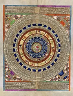
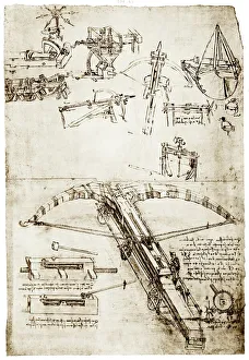
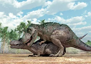
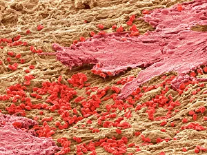
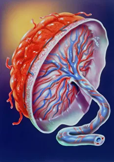
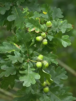
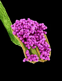
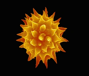
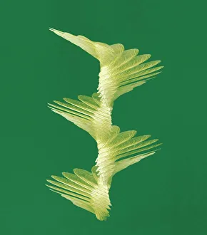
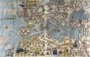
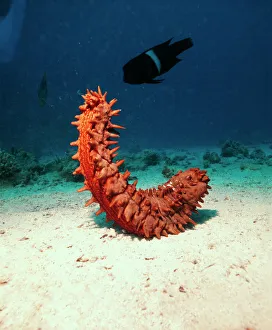
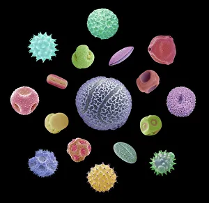
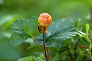
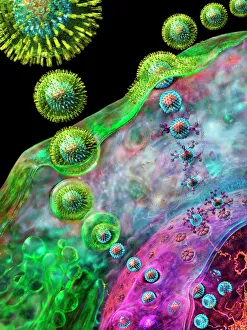
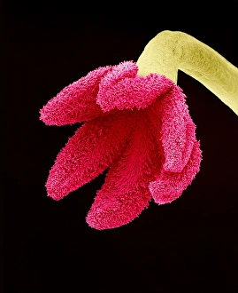
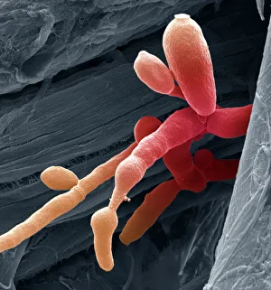
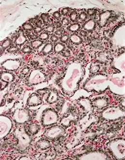
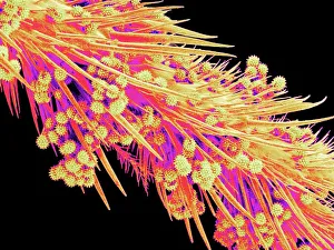
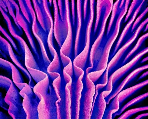
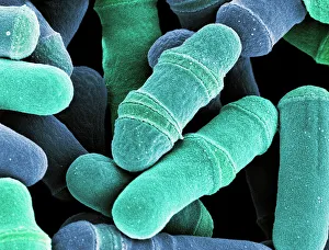
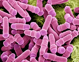
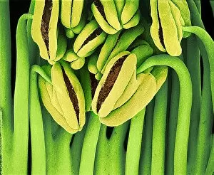
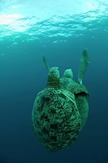
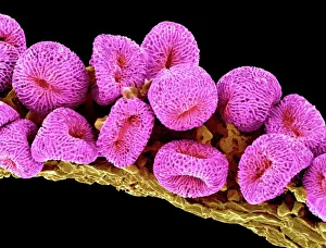
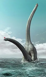
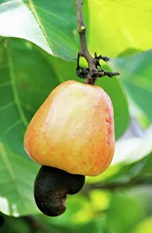
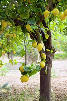
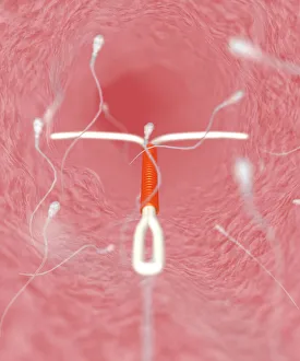
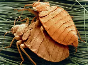
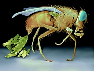
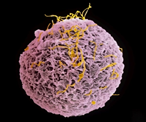
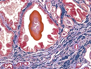
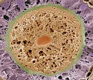
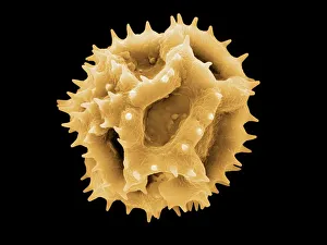
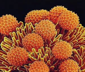
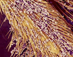
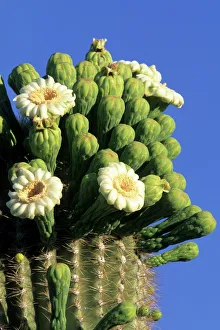
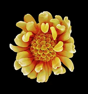
Exploring the wonders of reproduction through time and nature. 🌍🔬 From the intricate Catalan Atlas of the 14th century to Da Vinci's innovative crossbow, humans have always sought ways to understand and enhance reproduction. Witness the ancient mating rituals of Tyrannosaurus rex dinosaurs captured in fossils, or marvel at the microscopic beauty of a uterus lining during menstruation under an SEM microscope. Delve into the miracle of life with illustrations showcasing the human placenta or observe English oak acorns as they prepare for their own reproductive journey. Discover nature's secrets with stunning images of geranium anthers and dahlia flower pollen magnified by SEM technology. Follow maple seed flight paths as they embark on their quest for new beginnings, just like Robert Louis Stevenson once wrote about in his captivating works on reproduction. Dive deep into marine life with sea cucumbers, fascinating creatures that possess unique methods of reproduction. And finally, explore the intricate world of plant fertility with mesmerizing SEM images capturing pollen grains in all their glory. Join us on this incredible journey through time and nature as we unravel the mysteries behind 'reproduction'.