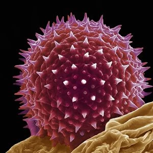
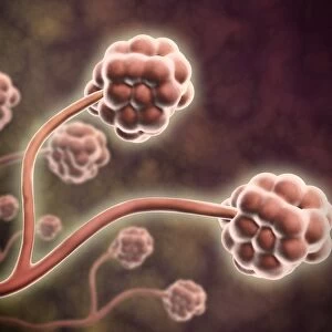
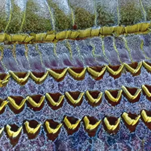
Coccolith. Scanning electron microscope (SEM) image of a Folkestone chalk
 image of a Folkestone chalk")
![]()
Wall Art and Photo Gifts from Mary Evans Picture Library
Coccolith. Scanning electron microscope (SEM) image of a Folkestone chalk
Scanning electron microscope (SEM) image of a Folkestone chalk surface with Cretaceous coccoliths (x2500 on a standard 9 cm wide print)
Mary Evans Picture Library makes available wonderful images created for people to enjoy over the centuries
Media ID 8591191
© Mary Evans Picture Library 2015 - https://copyrighthub.org/s0/hub1/creation/maryevans/MaryEvansPictureID/10708485
Abstract Alga Algae Algal Celled Chalk Chromalveolata Chromista Coccolith Coccolithophore Coccolithophorid Electron Micrograph Eukaryote Eukaryotic Haptophyta Haptophyte Magnification Micrograph Microscope Image Phytoplankton Plankton Planktonic Protist Protista Invertebrata
EDITORS COMMENTS
1. Title: Magnificent Coccolith Array: A Closer Look into the Microcosm of the Cretaceous Sea. This stunning Scanning Electron Microscope (SEM) image showcases the intricate and mesmerizing pattern of coccoliths, tiny calcium carbonate plates produced by the single-celled organisms known as coccolithophores. The image captures a surface of Folkestone chalk, a sedimentary rock formed during the Late Cretaceous period, teeming with these microscopic algae. Coccolithophores are a crucial part of the marine plankton community, belonging to the phylum Haptophyta within the kingdom Chromista. These eukaryotic organisms, classified as protists, are responsible for the production of approximately 50% of the calcium carbonate deposited in the world's oceans. The coccoliths, shaped like tiny discs or plates, protect these organisms and provide them with buoyancy, enabling them to float in the water column. The intricate patterns on the plates vary between different species, making them valuable tools for paleontologists and geologists in studying the past climate and ocean conditions. The image, taken at a magnification of x2500, reveals the abstract beauty of these microscopic organisms, their complex cellular structures, and the intricate patterns of their coccoliths. This photograph serves as a reminder of the diverse and intricate world that exists beyond our naked eyes, inviting us to explore the wonders of the natural world and the secrets it holds. The Cretaceous period, marked by the dominance of these microscopic organisms, ended approximately 66 million years ago, leaving behind a rich fossil record that continues to reveal new insights into the history of life on Earth. This image is a testament to the beauty and importance of the smallest organisms in our oceans and the role they play in shaping our planet.
MADE IN THE USA
Safe Shipping with 30 Day Money Back Guarantee
FREE PERSONALISATION*
We are proud to offer a range of customisation features including Personalised Captions, Color Filters and Picture Zoom Tools
FREE COLORIZATION SERVICE
You can choose advanced AI Colorization for this picture at no extra charge!
SECURE PAYMENTS
We happily accept a wide range of payment options so you can pay for the things you need in the way that is most convenient for you
* Options may vary by product and licensing agreement. Zoomed Pictures can be adjusted in the Cart.