
Eukaryotic Collection
Eukaryotic organisms encompass a vast array of life forms, ranging from the microscopic to the towering kelp forests in our oceans


All Professionally Made to Order for Quick Shipping
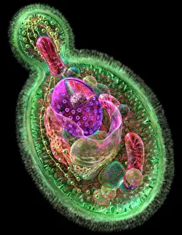
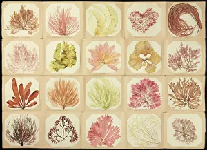
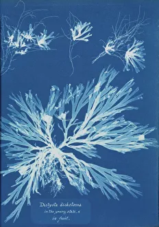
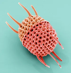
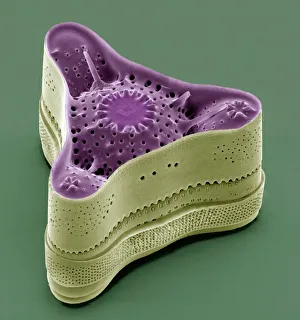
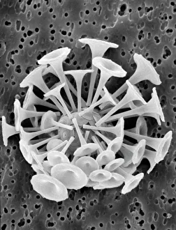
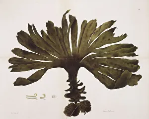
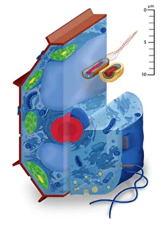
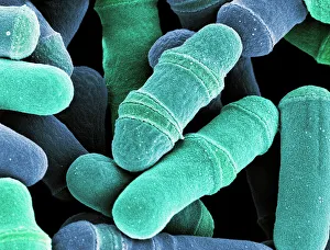
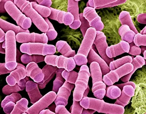
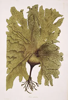
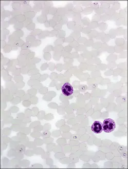
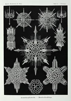
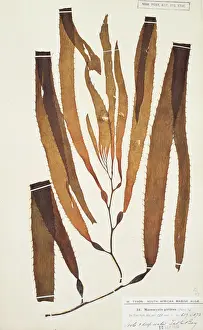
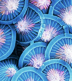
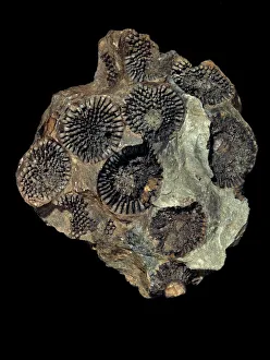
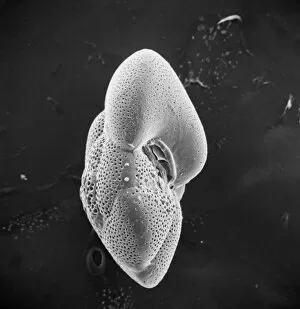
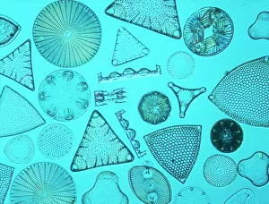
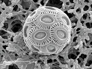
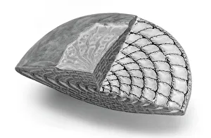
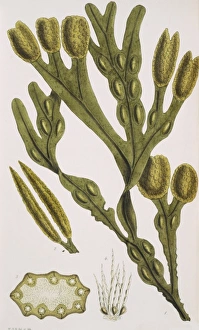
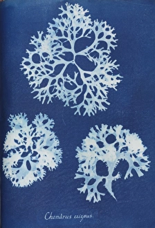
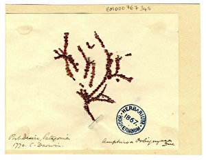
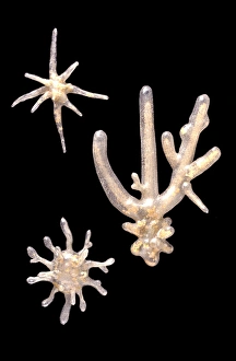
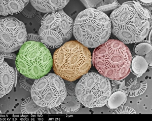
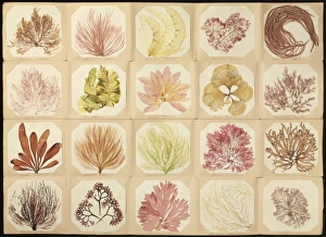
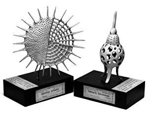
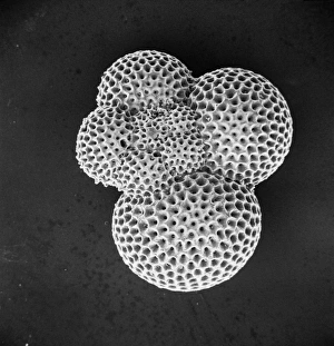
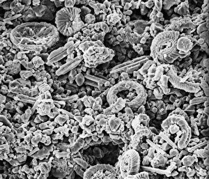
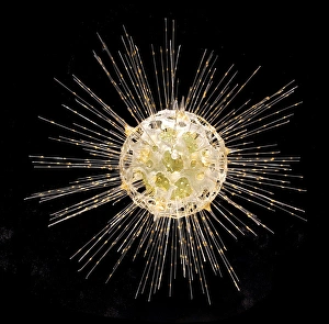
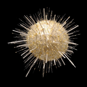
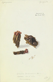
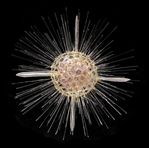
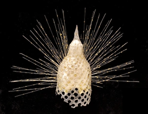
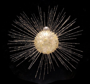
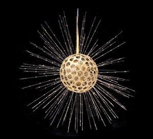
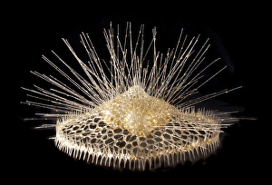
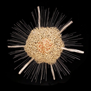
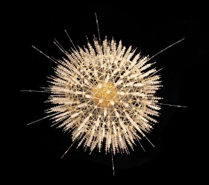
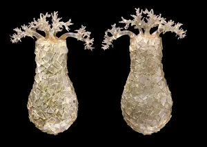
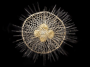
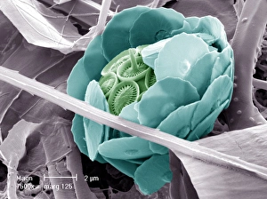
Eukaryotic organisms encompass a vast array of life forms, ranging from the microscopic to the towering kelp forests in our oceans. Take a closer look at these captivating hints that showcase the diversity and complexity of eukaryotes. Starting with budding yeast cells, we witness their remarkable ability to reproduce through cell division. Under the scanning electron microscope (SEM), dividing yeast cells reveal intricate structures and processes that contribute to their survival and proliferation. Moving on to pressed seaweed specimens, such as Dictyota dichotoma and Fucus bulbosus, we explore the fascinating world of marine algae. These eukaryotic organisms play crucial roles in ocean ecosystems by providing shelter for countless other species while also contributing to nutrient cycling. Delving deeper into microscopic wonders, we encounter Diatoms under SEM. These single-celled eukaryotes exhibit stunning geometric patterns on their silica-based shells. Their ecological significance cannot be overstated as they are responsible for a significant portion of Earth's oxygen production. Another mesmerizing microorganism is Discosphaera tubifera, commonly known as coccolithophore. These tiny calcifying algae adorn themselves with intricately sculpted calcium carbonate plates called coccoliths. Their presence can create breathtaking blooms visible from space. Shifting gears towards Plasmodium sp. , an insidious malarial parasite that infects human red blood cells, we confront one of the darker aspects lifeforms. This reminder highlights how even within this kingdom there exists both beauty and danger. To round out our exploration, let's not forget about majestic kelps like Fucus radiatus. These large brown algae form underwater forests teeming with biodiversity while serving as vital carbon sinks in our changing climate. Finally, artistic renditions depicting various cell types remind us that behind every scientific discovery lies creativity and imagination – essential tools for unraveling nature's mysteries.