
Algal Collection
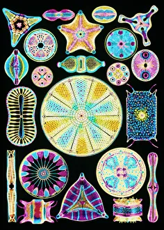
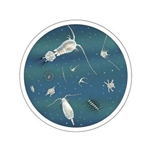
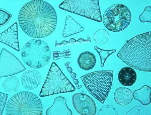
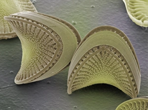
"Exploring the Intricate World Beauty" Immerse yourself in the captivating artistry of diatom algae, as depicted by Ernst Haeckel


All Professionally Made to Order for Quick Shipping
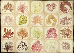
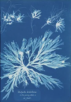
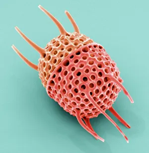
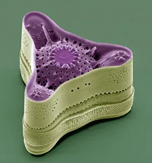
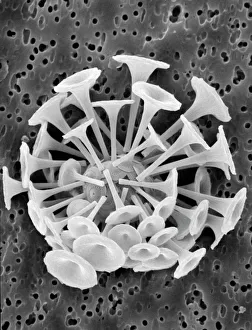
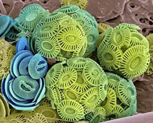
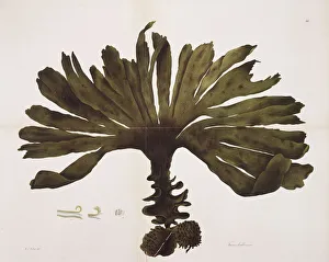
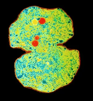
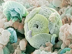
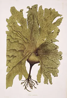
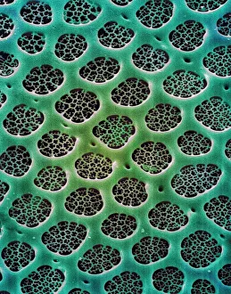
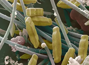
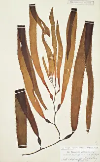
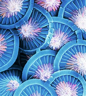
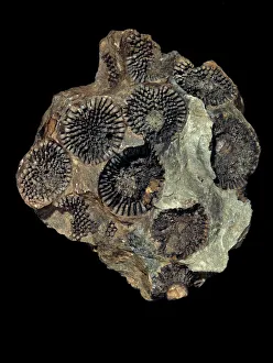
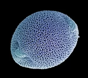
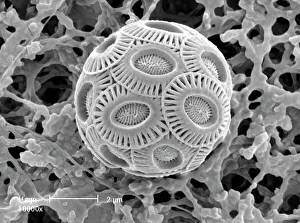
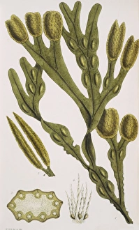
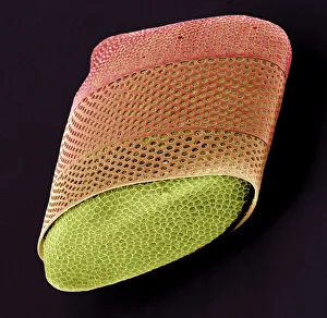
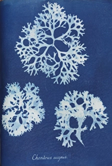
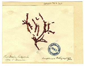
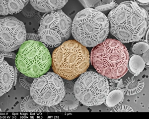
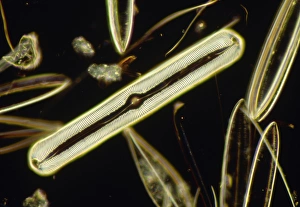
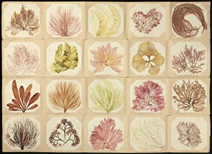
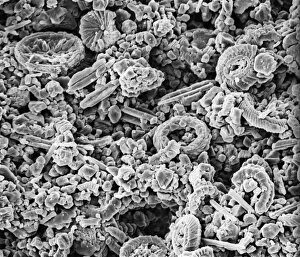
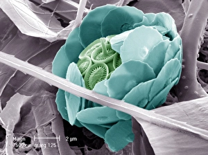
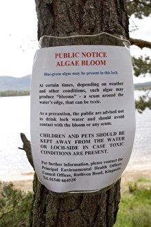
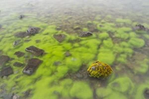
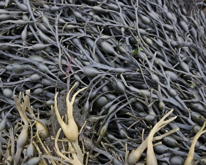
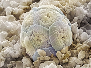
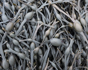
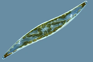
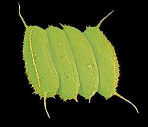
"Exploring the Intricate World Beauty" Immerse yourself in the captivating artistry of diatom algae, as depicted by Ernst Haeckel. These microscopic organisms showcase their intricate structures and mesmerizing patterns, reminding us of the boundless creativity found in nature. Pressed seaweed specimens C016 / 6127 offer a glimpse into the diverse forms and textures that algal species can take. From delicate filaments to robust fronds, each specimen tells a unique story of adaptation and survival in marine ecosystems. Behold the beauty of calcareous phytoplankton through scanning electron microscopy (SEM). The exquisite details captured reveal their ornate shells and elaborate architecture, serving as a testament to their vital role in oceanic food webs. Discosphaera tubifera, a coccolithophore, enchants with its spherical shape adorned by intricate calcium carbonate plates. This tiny organism plays an essential part in carbon cycling and contributes to the stunning white cliffs seen along some coastlines. Dictyota dichotoma showcases its elegant branching structure, exemplifying how algae can create complex habitats for other marine organisms. Its presence enriches coastal ecosystems while providing shelter for countless creatures. Diatoms take center stage once again under SEM's magnifying lens. Their symmetrical siliceous skeletons exhibit remarkable diversity – from star-shaped patterns to delicate lace-like designs – showcasing nature's endless ingenuity. Marvel at Fucus bulbosus or kelp's majestic presence as it sways gracefully beneath ocean currents. These large brown algae provide refuge for numerous marine species while contributing to nutrient cycling within coastal environments. Microcystis blue-green alga captivates with its vibrant hue amidst freshwater bodies. Though often associated with harmful algal blooms, this organism also serves ecological functions such as nitrogen fixation and oxygen production. Witness yet another diatom masterpiece through SEM imagery – their intricately sculpted shapes and delicate ornamentation never cease to amaze.