Jigsaw Puzzle > Popular Themes > Human Body
Jigsaw Puzzle : Fractured ankle, X-ray


![]()
Jigsaw Puzzles from Science Photo Library
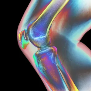
Fractured ankle, X-ray
Fractured ankle. Coloured profile X-ray of a distal fibula fracture (upper centre). The fibula is the smaller leg bone running down from top centre. The larger bone is the tibia. The ankle is the joint at which these bones meet the bones of the foot (lower right)
Science Photo Library features Science and Medical images including photos and illustrations
Media ID 6390143
© MIRIAM MASLO/SCIENCE PHOTO LIBRARY
Ankle Bones Broken Diagnosis Diagnostic Fibula Foot Fracture Fractured Injured Injury Joint Osteological Osteology Radiography Tibia X Ray X Ray Machine Condition Disorder False Coloured
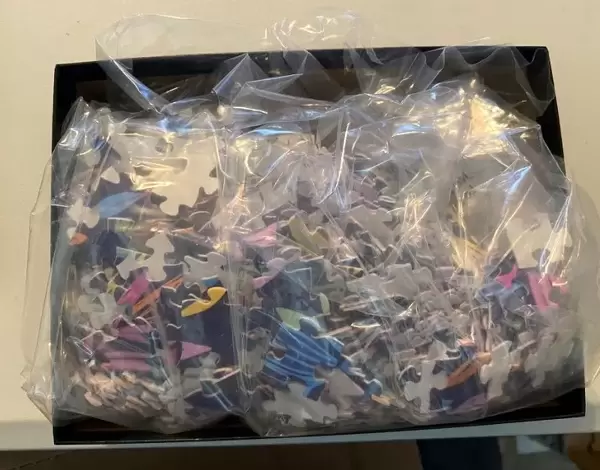
Jigsaw Puzzle (520 Pieces)
Discover the intricacies of the human body with Media Storehouse's Jigsaw Puzzles. Our latest addition to the collection showcases a fascinating X-ray image of a distal fibula fracture, captured by Science Photo Library. Piece together the vivid colors and intricate details of this coloured profile X-ray, revealing the complex structure of the ankle and the fracture in the smaller fibula bone. A challenging and educational puzzle for adults and older children alike, this engaging activity will keep you hooked until the final piece falls into place. Explore the wonders of science and anatomy with Media Storehouse's Jigsaw Puzzles.
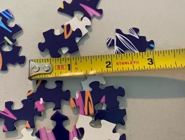
Made in the USA, 520-piece puzzles measure 16" x 20" (40.6 x 50.8 cm). Every puzzle is meticulously printed on glossy photo paper, which has a strong 1.33 mm thickness. Delivered in a black storage cardboard box, these puzzles are both stylish and practical. (Note: puzzles contain small parts and are not suitable for children under 3 years of age.)
Jigsaw Puzzles are an ideal gift for any occasion
Estimated Product Size is 40.5cm x 50.8cm (15.9" x 20")
These are individually made so all sizes are approximate
Artwork printed orientated as per the preview above, with landscape (horizontal) or portrait (vertical) orientation to match the source image.
EDITORS COMMENTS
This print from Science Photo Library showcases a visually striking representation of a fractured ankle. The image, taken using an X-ray machine, reveals the intricate details of this injury in vivid color. At the upper center, we can clearly observe a distal fibula fracture - one of the two bones that make up our lower leg. The larger bone adjacent to it is the tibia, which forms part of the ankle joint. The ankle joint itself is prominently displayed at the lower right corner, where these crucial bones meet with those of the foot. This diagnostic tool provides valuable insight into understanding and diagnosing such conditions accurately. The false-colored hues employed in this photograph enhance its visual impact while maintaining scientific accuracy. It serves as a powerful reminder of both the fragility and resilience of our human bodies. This image not only appeals to medical professionals seeking to deepen their knowledge but also captivates anyone fascinated by anatomy and osteology. Its detailed portrayal offers an opportunity for individuals to appreciate how injuries like fractures can affect our mobility and overall well-being. As always, Science Photo Library delivers yet another remarkable piece that merges science with artistry flawlessly – capturing both educational value and aesthetic appeal within a single frame.
MADE IN THE USA
Safe Shipping with 30 Day Money Back Guarantee
FREE PERSONALISATION*
We are proud to offer a range of customisation features including Personalised Captions, Color Filters and Picture Zoom Tools
SECURE PAYMENTS
We happily accept a wide range of payment options so you can pay for the things you need in the way that is most convenient for you
* Options may vary by product and licensing agreement. Zoomed Pictures can be adjusted in the Cart.