Jigsaw Puzzle : Eye anatomy, SEM


![]()
Jigsaw Puzzles from Science Photo Library
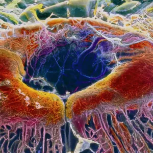
Eye anatomy, SEM
Eye anatomy. Coloured scanning electron micrograph (SEM) showing part of the ciliary body (blue) and iris (right) of an eye. The ciliary body is a ring-shaped structure that surrounds the iris and joins to ligaments that hold the lens in place behind the iris. It also contains the ciliary muscle that is contracted to alter the curvature of the lens and focus light on the retina at the back of the eye. Magnification: x20 when printed 10 centimetres wide
Science Photo Library features Science and Medical images including photos and illustrations
Media ID 6338269
© STEVE GSCHMEISSNER/SCIENCE PHOTO LIBRARY
Ciliary Body Colored False Colored Inside Internal Iris Ligament Ligaments Muscles Muscular Ocular Ophtalmological Ophthalmology Physiological Physiology Sight Tissue Vision False Coloured
Jigsaw Puzzle (520 Pieces)
Discover the intricacies of the human body with our Media Storehouse Jigsaw Puzzles! This captivating puzzle features a stunning Colored Scanning Electron Micrograph (SEM) image of the eye anatomy from Science Photo Library. Delve into the intricacies of the ciliary body in blue and the iris, gaining a new appreciation for the complexity of the eye. A perfect addition to any home or classroom, this jigsaw puzzle is not only a fun activity but also an educational experience. Engage your mind and challenge your problem-solving skills with this visually stimulating puzzle.
Made in the USA, 520-piece puzzles measure 16" x 20" (40.6 x 50.8 cm). Every puzzle is meticulously printed on glossy photo paper, which has a strong 1.33 mm thickness. Delivered in a black storage cardboard box, these puzzles are both stylish and practical. (Note: puzzles contain small parts and are not suitable for children under 3 years of age.)
Jigsaw Puzzles are an ideal gift for any occasion
Estimated Product Size is 50.8cm x 40.5cm (20" x 15.9")
These are individually made so all sizes are approximate
Artwork printed orientated as per the preview above, with landscape (horizontal) or portrait (vertical) orientation to match the source image.
EDITORS COMMENTS
This print from Science Photo Library offers a mesmerizing glimpse into the intricate world of eye anatomy. Through the lens of a scanning electron microscope (SEM), we are presented with a false-colored image that showcases the internal structures responsible for our vision. At first glance, our attention is drawn to the vibrant blue hues representing the ciliary body, which encircles and connects to ligaments holding the lens in place behind it. This ring-shaped structure plays a crucial role in adjusting the curvature of the lens through its contracted ciliary muscle, allowing us to focus light onto the retina at the back of our eyes. The level of detail captured by this SEM image is truly remarkable. Every muscular fiber and tissue within this delicate organ is brought to life, highlighting both its anatomical and physiological significance. It serves as a testament to how intricately designed our bodies are. As we delve deeper into understanding human biology, images like these become invaluable tools for researchers and medical professionals alike. They provide insights into ophthalmology and ocular health while fueling advancements in sight-related treatments. Whether you have an appreciation for scientific marvels or simply seek aesthetic beauty, this print invites you on an enlightening journey inside one of nature's most extraordinary creations –the human eye.
MADE IN THE USA
Safe Shipping with 30 Day Money Back Guarantee
FREE PERSONALISATION*
We are proud to offer a range of customisation features including Personalised Captions, Color Filters and Picture Zoom Tools
SECURE PAYMENTS
We happily accept a wide range of payment options so you can pay for the things you need in the way that is most convenient for you
* Options may vary by product and licensing agreement. Zoomed Pictures can be adjusted in the Cart.