Home > Science > SEM
Ciliary body

![]()
Wall Art and Photo Gifts from Science Photo Library
Ciliary body
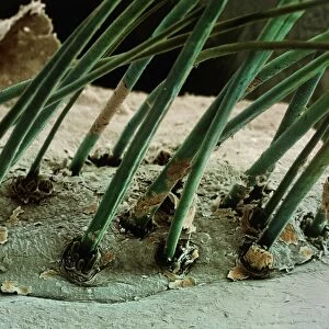
Ciliary body. Coloured scanning electron micrograph (SEM) of a section of the eye, showing the ciliary body and iris. The ciliary body (orange, lower left) forms a ring between the iris (green, upper right) and the choroid (the inner surface of the eyeball). The ciliary body joins to ligaments that hold the lens in place behind the iris. The ciliary body also contains the ciliary muscle that is contracted to alter the curvature of the lens and focus light on the retina
Science Photo Library features Science and Medical images including photos and illustrations
Media ID 6448999
© SUSUMU NISHINAGA/SCIENCE PHOTO LIBRARY
Ciliary Body False Colour Iris Optic Optics Sense Sight Vision False Coloured Section Sectioned
EDITORS COMMENTS
This print showcases the intricate beauty of the ciliary body, a vital component of our visual system. In this coloured scanning electron micrograph (SEM), we are granted an up-close view of a section of the eye, where the ciliary body takes center stage. Coloured in vibrant orange and situated at the lower left corner, the ciliary body forms a remarkable ring between two other prominent structures: the iris and the choroid. The iris, depicted in lush green hues at the upper right corner, is responsible for regulating light entering our eyes through its adjustable aperture. What makes this image even more fascinating is that it reveals how these anatomical elements work together seamlessly to ensure optimal vision. The ciliary body not only acts as a support structure but also houses an essential muscle known as the ciliary muscle. This muscle contracts or relaxes to modify lens curvature, enabling us to focus on objects at varying distances. As we delve deeper into understanding ocular anatomy through this SEM masterpiece by Science Photo Library, we gain insight into how our eyes function with precision and efficiency. It serves as a reminder of both nature's complexity and its ability to create wonders that allow us to perceive and appreciate all that surrounds us visually.
MADE IN THE USA
Safe Shipping with 30 Day Money Back Guarantee
FREE PERSONALISATION*
We are proud to offer a range of customisation features including Personalised Captions, Color Filters and Picture Zoom Tools
SECURE PAYMENTS
We happily accept a wide range of payment options so you can pay for the things you need in the way that is most convenient for you
* Options may vary by product and licensing agreement. Zoomed Pictures can be adjusted in the Cart.