Fraud Blocker Skip to main content

False Colored Collection

"Unveiling Nature's Hidden Palette: Exploring the World Microscopy" Step into a mesmerizing realm where science and art converge

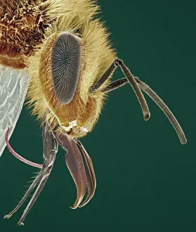
Background imageFalse Colored Collection: Head of a honey bee, SEM

Head of a honey bee, SEM
Head of a honey bee (Apis mellifera), coloured scanning electron micrograph (SEM). The compound eyes (one seen) are either side of the antennae

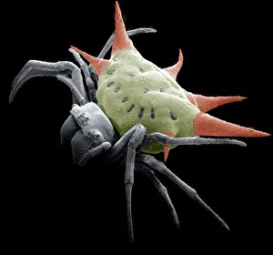
Background imageFalse Colored Collection: Spiny spider, SEM

Spiny spider, SEM
Spiny spider. Coloured scanning electron micrograph (SEM) of a spiny spider (Gasteracantha sp.), showing its characteristic spines (red) and large mouthparts (grey, centre left)

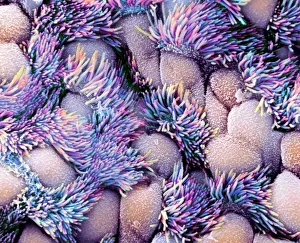
Background imageFalse Colored Collection: Trachea lining, SEM

Trachea lining, SEM
Trachea lining, coloured scanning electron micrograph (SEM). The tracheal lining contains mucus-secreting goblet cells and epithelial cells bearing hair-like cilia (blue)

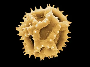
Background imageFalse Colored Collection: Dandelion pollen grain, SEM

Dandelion pollen grain, SEM
Dandelion (Taraxacum officinale) pollen grain, coloured scanning electron micrograph (SEM). Pollen grains are reproductive structures produced by the male parts of flowering plants

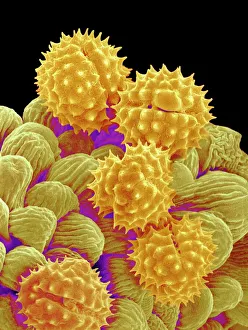
Background imageFalse Colored Collection: Philadelphia fleabane pollen grains, SEM

Philadelphia fleabane pollen grains, SEM
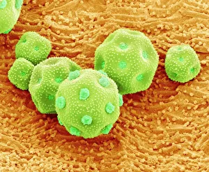
Philadelphia fleabane (Erigeron philadelphicus) pollen grains, coloured scanning electron micrograph (SEM). Pollen grains are reproductive structures produced by the male parts of flowering plants

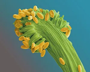
Background imageFalse Colored Collection: Gorse stigma with pollen grains, SEM

Gorse stigma with pollen grains, SEM
Gorse (Ulex europaeus) stigma (green) with pollen grains (yellow), coloured scanning electron micrograph (SEM). Pollen grains are reproductive structures produced by the male parts of flowering

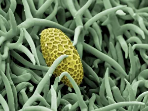
Background imageFalse Colored Collection: Lily pollen grain on rosemary leaf, SEM

Lily pollen grain on rosemary leaf, SEM
Lily pollen grain on rosemary leaf. Coloured scanning electron micrograph (SEM) of a pollen grain (yellow) from a lily flower (Lilium sp.)

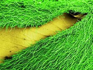
Background imageFalse Colored Collection: Gorse flower bud, SEM

Gorse flower bud, SEM
Gorse (Ulex europaeus) flower bud, coloured scanning electron micrograph (SEM). One of the petals (yellow) is seen enclosed in the flower casing (calyx)

Background imageFalse Colored Collection: Forsythia pollen grains, SEM

Forsythia pollen grains, SEM
Forsythia pollen grains arranged in a spiral, coloured scanning electron micrograph (SEM). Pollen grains are reproductive structures produced by the male parts of flowering plants

Background imageFalse Colored Collection: Chickweed pollen grains, SEM

Chickweed pollen grains, SEM
Chickweed pollen grains. Coloured scanning electron micrograph (SEM) of pollen grains (green) from a chickweed flower (Stellaria media)

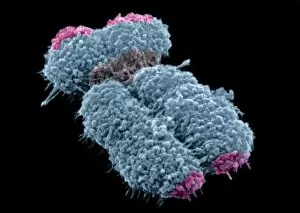
Background imageFalse Colored Collection: Human chromosomes, SEM C013 / 5002

Human chromosomes, SEM C013 / 5002
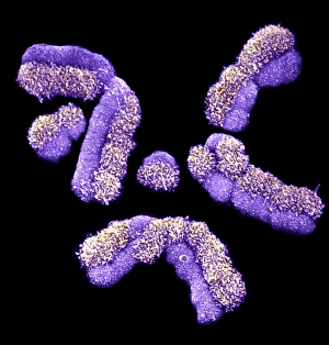
Human chromosomes. Coloured scanning electron micrograph (SEM) of human chromosomes prepared with the harlequin staining technique

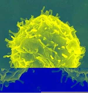
Background imageFalse Colored Collection: Haematopoietic stem cells, SEM C013 / 5009

Haematopoietic stem cells, SEM C013 / 5009
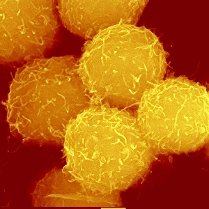
Haematopoietic stem cells, coloured scanning electron micrograph (SEM). Stem cells can differentiate into any other cell type

Background imageFalse Colored Collection: Osteoblast bone cell, SEM

Osteoblast bone cell, SEM
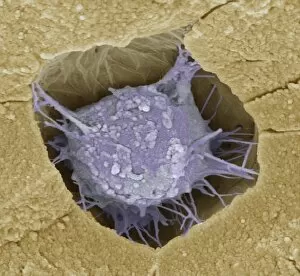
Osteoblast bone cell. Coloured scanning electron micrograph (SEM) of an osteoblast (centre) in bone tissue. Osteoblasts are primarily found in regions of new bone growth

Background imageFalse Colored Collection: Chickweed pollen grain, SEM

Chickweed pollen grain, SEM
Chickweed pollen grain. Coloured scanning electron micrograph (SEM) of a pollen grain (green) from a chickweed flower (Stellaria media)

Background imageFalse Colored Collection: Honey bee head, SEM

Honey bee head, SEM
Honey bee head. Coloured scanning electron micrograph (SEM) of the head of a honey bee (Apis sp.). The compound eyes (red) are either side of the antennae
Background imageFalse Colored Collection: Human chromosome, SEM C013 / 4999

Human chromosome, SEM C013 / 4999
Human chromosome. Coloured scanning electron micrograph (SEM) of a human chromosome. Chromosomes are a packaged form of a cells genetic material DNA (deoxyribonucleic acid)

Background imageFalse Colored Collection: Prostate cancer cell, SEM C013 / 5012

Prostate cancer cell, SEM C013 / 5012
Prostate cancer cell, coloured scanning electron micrograph (SEM). The uneven surface of this cell is typical of cancer cells

Background imageFalse Colored Collection: Prostate cancer cell, SEM C013 / 5011

Prostate cancer cell, SEM C013 / 5011
Prostate cancer cell, coloured scanning electron micrograph (SEM). The uneven surface of this cell is typical of cancer cells

Background imageFalse Colored Collection: Haematopoietic stem cell, SEM C013 / 5008

Haematopoietic stem cell, SEM C013 / 5008
Haematopoietic stem cell, coloured scanning electron micrograph (SEM). Stem cells can differentiate into any other cell type

Background imageFalse Colored Collection: Haematopoietic stem cell, SEM C013 / 5007

Haematopoietic stem cell, SEM C013 / 5007
Haematopoietic stem cell, coloured scanning electron micrograph (SEM). Stem cells can differentiate into any other cell type

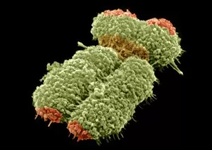
Background imageFalse Colored Collection: Human chromosomes, SEM C013 / 5005

Human chromosomes, SEM C013 / 5005
Human chromosomes. Coloured scanning electron micrograph (SEM) of human chromosomes prepared with the harlequin staining technique

Background imageFalse Colored Collection: Haematopoietic stem cell, SEM C013 / 5006

Haematopoietic stem cell, SEM C013 / 5006
Haematopoietic stem cell, coloured scanning electron micrograph (SEM). Stem cells can differentiate into any other cell type

Background imageFalse Colored Collection: Human chromosome, SEM C013 / 4998

Human chromosome, SEM C013 / 4998
Human chromosome. Coloured scanning electron micrograph (SEM) of a human chromosome. Chromosomes are a packaged form of a cells genetic material DNA (deoxyribonucleic acid)

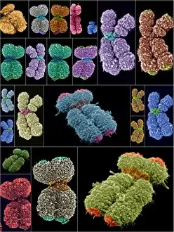
Background imageFalse Colored Collection: Human chromosomes, SEMs

Human chromosomes, SEMs
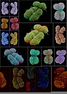
Human chromosomes. Collage of coloured scanning electron micrographs (SEMs) of human chromosomes. Chromosomes are a packaged form of a cells genetic material DNA (deoxyribonucleic acid)

Background imageFalse Colored Collection: Human chromosomes, SEMs C013 / 4989

Human chromosomes, SEMs C013 / 4989
Human chromosomes. Collage of coloured scanning electron micrographs (SEMs) of human chromosomes. Chromosomes are a packaged form of a cells genetic material DNA (deoxyribonucleic acid)

Background imageFalse Colored Collection: Primate finger bone, SEM

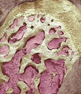
Primate finger bone, SEM

Background imageFalse Colored Collection: Primate fingerprint ridges, SEM

Primate fingerprint ridges, SEM

Background imageFalse Colored Collection: Primate eyelash, SEM

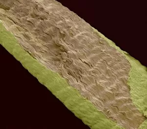
Primate eyelash, SEM

Background imageFalse Colored Collection: Squirrel monkey incisors, SEM

Squirrel monkey incisors, SEM

Background imageFalse Colored Collection: Primate finger muscle, SEM

Primate finger muscle, SEM

Background imageFalse Colored Collection: Stem cells, light micrograph

Stem cells, light micrograph
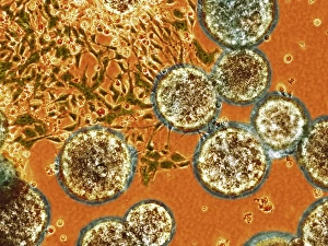
Stem cells. Coloured light micrograph of stem cells undergoing spontaneous differentiation. Stem cells are precursor cells that can differentiate spontaneously or in a directed fashion to form

Background imageFalse Colored Collection: Primate ear canal, SEM

Primate ear canal, SEM

Background imageFalse Colored Collection: Primate hand tendon, SEM

Primate hand tendon, SEM

Background imageFalse Colored Collection: Water spider, SEM

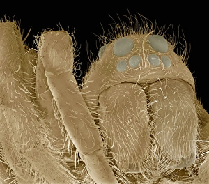
Water spider, SEM
Water spider. Coloured scanning electron micrograph (SEM) of the head of a water spider (Argyroneta aquatica), showing its large jaws (mandibles, centre right) and its eyes (upper right). A

Background imageFalse Colored Collection: Frog skin, SEM

Frog skin, SEM
Frog skin. Coloured scanning electron micrograph (SEM) of the skin of a frog, showing numerous folds (microplicae). These folds maximise the surface area to allow for increased cutaneous gas

Background imageFalse Colored Collection: Frog pigment cell, SEM

Frog pigment cell, SEM
Frog pigment cell. Coloured scanning electron micrograph (SEM) of a section through a chromatophore pigment cell from the skin of a frog

Background imageFalse Colored Collection: Antlion larva, SEM

Antlion larva, SEM
Antlion larva, coloured scanning electron micrograph (SEM). The larval form of this insect (family Myrmeleontide) lives at the bottom of a cone shaped hole

Background imageFalse Colored Collection: Mosquito head, SEM

Mosquito head, SEM
Mosquito head. Coloured scanning electron micrograph (SEM) of the head of a mosquito, showing its large compound eyes. Each eye consists of many lenses (spheres) known as ommatidia

Background imageFalse Colored Collection: Frogspawn, SEM

Frogspawn, SEM
Frogspawn. Coloured scanning electron micrograph (SEM) of a section through frog eggs, showing the large nucleus containing nucleoli, surrounded by yolk

Background imageFalse Colored Collection: Pancreas tissue, SEM

Pancreas tissue, SEM
Pancreas tissue. Coloured scanning electron micrograph (SEM) of fractured pancreas tissue, showing numerous acinar cells, containing secretory zymogen granules

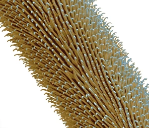
Background imageFalse Colored Collection: Tarantula hair, SEM

Tarantula hair, SEM
Tarantula hair. Coloured scanning electron micrograph (SEM) of a hair from the abdomen of a tarantula (family Theraphosidae) spider

Background imageFalse Colored Collection: Millipede head, SEM

Millipede head, SEM
Millipede head. Coloured scanning electron micrograph (SEM) of the head of a millipede from Barbados. Millipedes (class Diplopoda) have elongated bodies with two pairs of legs on each body segment

Background imageFalse Colored Collection: Spinal cord, SEM

Spinal cord, SEM
Spinal cord. Coloured scanning electron micrograph (SEM) of a section through a spinal cord, showing the grey matter (green) and white matter (blue)

Background imageFalse Colored Collection: Middle ear bone, SEM

Middle ear bone, SEM
Middle ear bone. Coloured scanning electron micrograph (SEM) of the human middle ear, showing the stapes (ring-shaped). The stapes is one of three bones (known as the ossicles)

Background imageFalse Colored Collection: Retina blood vessel, SEM

Retina blood vessel, SEM
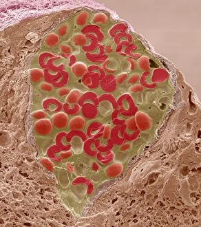
Retina blood vessel. Coloured scanning electron micrograph (SEM) of a section through a blood vessel in the retina of an eye, showing the red blood cells (red)

Background imageFalse Colored Collection: Iris pigment epithelium, SEM

Iris pigment epithelium, SEM
Iris pigment epithelium. Coloured scanning electron micrograph (SEM) of a section through the iris of an eye, showing the iris pigment epithelium (IPE)

Background imageFalse Colored Collection: Booklouse, SEM

Booklouse, SEM
Booklouse. Coloured scanning electron micrograph (SEM) of the head of a booklouse (order Psocoptera). These insects, also known as barklice or barkflies

Background imageFalse Colored Collection: Optic nerve fibres, SEM

Optic nerve fibres, SEM
Optic nerve fibres. Coloured scanning electron micrograph (SEM) of axons from the optic nerve. Connective tissue fibres (thread-like) are also visible


All Professionally Made to Order for Quick Shipping

"Unveiling Nature's Hidden Palette: Exploring the World Microscopy" Step into a mesmerizing realm where science and art converge, revealing captivating details that are invisible to the naked eye. In this collection images captured through scanning electron microscopy (SEM), we embark on a journey through various specimens, each showcasing their unique beauty. First up, we encounter the intricate head of a honey bee, magnified to expose its delicate features in stunning hues. Moving on, we delve into the spiny world of a spider as its exoskeleton is brought to life with vibrant colors under SEM. Venturing deeper into nature's wonders, we explore the trachea lining - an enchanting network resembling an otherworldly landscape. Next, dandelion pollen grains take center stage; their textured surfaces transformed by false coloring techniques into an ethereal spectacle. As our exploration continues, Philadelphia fleabane pollen grains come alive in vivid shades against a dark backdrop. The gorse stigma adorned with pollen grains follows suit – an exquisite display reminiscent of abstract art. A surprising juxtaposition awaits as we discover a lily pollen grain delicately resting upon a rosemary leaf – contrasting textures and colors intertwining harmoniously. Meanwhile, the gorse flower bud reveals hidden secrets within its petals when observed at microscopic levels. Nature's diversity unfolds further with Forsythia pollen grains taking shape in brilliant tones that evoke feelings of warmth and vitality. Chickweed pollen grains add another layer to this symphony of color and form – tiny spheres bursting forth like miniature fireworks frozen in time. Intriguingly shifting gears from flora to fauna, human chromosomes emerge as striking patterns under SEM C013 / 5002 – reminding us how intricately woven our genetic makeup truly is. Finally, optic nerve fibers weave together like cosmic threads connecting our visual perception to reality itself. Through these false colored masterpieces created by scientific exploration, we are reminded of the hidden beauty that lies within our world.