Fraud Blocker Skip to main content

Tissue Collection (page 9)

"Tissue: A Microscopic World Unveiled" Uterus lining during menstruation, SEM: Witness the intricate dance of life as the uterus sheds its lining during menstruation

Background imageTissue Collection: Parkinsons disease, light micrograph

Parkinsons disease, light micrograph
Parkinsons disease. Light micrograph of a section through the sabstantia nigra of the brain of a patient with Parkinsons disease showing a neuron (nerve cell)

Background imageTissue Collection: Dementia, light micrograph

Dementia, light micrograph
Dementia. Light micrograph of a section through the brain of a patient with dementia. Dementia is mainly a disease of old age

Background imageTissue Collection: Ductal breast cancer, TEM

Ductal breast cancer, TEM
Ductal breast cancer. Transmission electron micrograph (TEM) of a section through breast tissue, showing malignant (cancerous) epithelial cells arising from the milk ducts of the breast

Background imageTissue Collection: Corpus luteum, light micrograph

Corpus luteum, light micrograph
Corpus luteum. Light micrograph of a section through the corpus luteum of an ovary. The corpus luteum is the tissue formed from a mature ovarian follicle after the follicle has released its oocyte

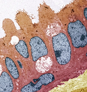
Background imageTissue Collection: Smell receptor, TEM

Smell receptor, TEM
Smell receptor. Transmission electron micrograph (TEM) of a section through the olfactory epithelium that lines the nasal cavity, showing an olfactory cell (smell receptor)

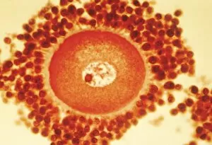
Background imageTissue Collection: Oocyte before ovulation, light micrograph

Oocyte before ovulation, light micrograph
Oocyte before ovulation. Light micrograph of a section through tissue from an ovary, showing an oocyte two days before ovulation

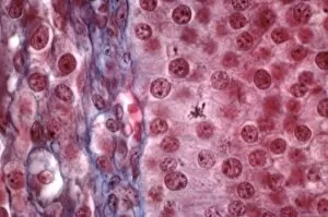
Background imageTissue Collection: Ovarian follicle tissue, light micrograph

Ovarian follicle tissue, light micrograph
Ovarian follicle tissue. Light micrograph of a section through tissue from an ovarian follicle, showing a boundary between two layers

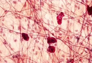
Background imageTissue Collection: Mast cells, light micrograph

Mast cells, light micrograph
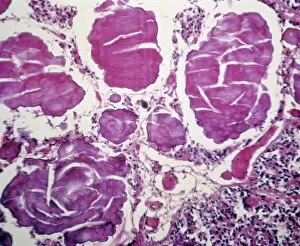
Mast cells. Light micrograph of several mast cells (pink rounded objects). These leucocytes (white blood cells) contain the chemical mediators histamine, serotonin and heparin

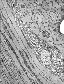
Background imageTissue Collection: Smooth muscle of gut wall, TEM

Smooth muscle of gut wall, TEM
Smooth muscle of gut wall. Transmission electron micrograph (TEM) of a section through the outermost wall of the intestine

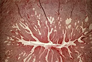
Background imageTissue Collection: Cervix, light micrograph

Cervix, light micrograph
Cervix. Light micrograph of a section through the mucosa of the cervix, the neck of the uterus. This is the endocervix, which borders the uterus. It is lined with columnar epithelial cells

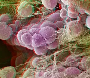
Background imageTissue Collection: Fat tissue, stereoscopic SEM

Fat tissue, stereoscopic SEM
Fat tissue. Coloured stereoscopic (3D) scanning electron micrograph (SEM) of a sample of fat tissue, showing fat cells (adipocytes, round) surrounded by fine strands of supportive connective tissue

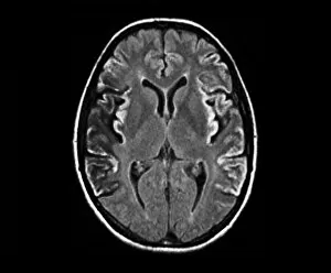
Background imageTissue Collection: Creutzfeldt-Jakob disease, MRI scan

Creutzfeldt-Jakob disease, MRI scan
Creutzfeldt-Jakob disease. Coloured magnetic resonance imaging (MRI) scan of an axial section through the brain of a 35-year-old patient

Background imageTissue Collection: Pineal gland, light micrograph

Pineal gland, light micrograph. The pineal gland is a small endocrine gland located near to the centre of the brain, between the two hemispheres

Background imageTissue Collection: Kidney tumour, CT scan

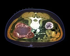
Kidney tumour, CT scan
Kidney tumour. Coloured computed tomography (CT) scan of a section through the abdomen of a 62-year-old patient, showing necrosis (death) of tissue in the right kidney

Background imageTissue Collection: Smooth muscle tissue, TEM

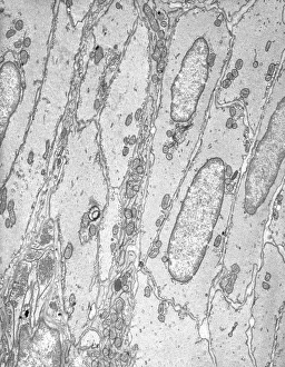
Smooth muscle tissue, TEM
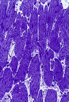
Smooth muscle tissue. Transmission electron micrograph (TEM) of a longitudinal section through smooth muscle cells. Smooth muscle cells are spindle-shaped and often closely packed together

Background imageTissue Collection: Thyroid follicles, light micrograph

Thyroid follicles, light micrograph
Thyroid follicles. Light micrograph of a section through a thyroid gland showing the follicles. The follicles are lined by a single layer of cuboidal epithelial cells

Background imageTissue Collection: Pancreas cells, SEM C015 / 9502

Pancreas cells, SEM C015 / 9502
Pancreas cells, coloured scanning electron micrograph (SEM). The small sections seen here are clusters of acini cells. These are exocrine cells, secreting digestive enzymes

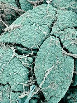
Background imageTissue Collection: Pancreas lobes, SEM C015 / 9496

Pancreas lobes, SEM C015 / 9496
Pancreas lobes, coloured scanning electron micrograph (SEM). Several lobes of the pancreas are seen here, separated by fissures. The smaller sections seen on each lobe are clusters of acini cells

Background imageTissue Collection: Heart tissue, SEM C015 / 9598

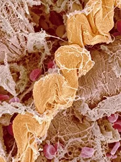
Heart tissue, SEM C015 / 9598
Heart tissue. Coloured scanning electron micrograph (SEM) of heart tissue with red blood cells (erythrocytes, red) and connective tissue (orange)

Background imageTissue Collection: Gastritis, light micrograph C016 / 0508

Gastritis, light micrograph C016 / 0508
Gastritis. Light micrograph of a section through an inflamed human stomach (gastritis), showing an abnormal accumulation of white blood cells (leucocytes)

Background imageTissue Collection: Liver, SEM C015 / 9924

Liver, SEM C015 / 9924
Liver vein. Coloured scanning electron micrograph (SEM) of liver tissue. The functions of the liver include destroying old red blood cells

Background imageTissue Collection: Corpus luteum of ovary, light micrograph C016 / 0527

Corpus luteum of ovary, light micrograph C016 / 0527
Corpus luteum of ovary. Light micrograph of a section through steroid-secreting luteal cells in the corpus luteum of an ovary. The corpus luteum develops from an ovulated follicle

Background imageTissue Collection: Pancreas lobes, SEM C015 / 9498

Pancreas lobes, SEM C015 / 9498
Pancreas lobes, coloured scanning electron micrograph (SEM). Several lobes of the pancreas are seen here, separated by fissures. The smaller sections seen on each lobe are clusters of acini cells

Background imageTissue Collection: Appendix, light micrograph C016 / 0510

Appendix, light micrograph C016 / 0510
Appendix. Light micrograph of a section through a human appendix. The appendix is a blind-ending small diameter tubular extension of the gut, arising from the caecum of the large bowel

Background imageTissue Collection: Retina of the eye, light micrograph C016 / 0528

Retina of the eye, light micrograph C016 / 0528
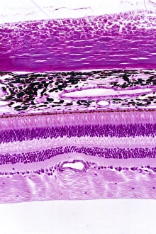
Retina of the eye. Light micrograph of a section through the retina from a human eye. From top down: nerve fibres of the optic nerve and a blood vessel; several layers of neurons (nerve cells)

Background imageTissue Collection: Gastritis, light micrograph C016 / 0507

Gastritis, light micrograph C016 / 0507
Gastritis. Light micrograph of a section through an inflamed human stomach (gastritis), showing an abnormal accumulation of white blood cells (leucocytes)

Background imageTissue Collection: Cancellous bone, light micrograph C016 / 0509

Cancellous bone, light micrograph C016 / 0509
Cancellous bone. Light micrograph of a section through cancellous, or spongy, bone. Constructed in a honeycomb-type architecture

Background imageTissue Collection: Pancreas cells, SEM C015 / 9501

Pancreas cells, SEM C015 / 9501
Pancreas cells, coloured scanning electron micrograph (SEM). The small sections seen here are clusters of acini cells. These are exocrine cells, secreting digestive enzymes

Background imageTissue Collection: Liver, SEM C015 / 9922

Liver, SEM C015 / 9922
Liver vein. Coloured scanning electron micrograph (SEM) of liver tissue showing a large vein (pink, up centre). The functions of the liver include destroying old red blood cells

Background imageTissue Collection: Pancreas lobes, SEM C015 / 9500

Pancreas lobes, SEM C015 / 9500
Pancreas lobes, coloured scanning electron micrograph (SEM). Several lobes of the pancreas are seen here, separated by fissures. The smaller sections seen on each lobe are clusters of acini cells

Background imageTissue Collection: Colon polyp, light micrograph C016 / 0512

Colon polyp, light micrograph C016 / 0512
Colon polyp. Light micrograph of a section through part of a tubulovillous adenoma of the mucosa of the large bowel (colon)

Background imageTissue Collection: Lens of the eye, light micrograph C016 / 0526

Lens of the eye, light micrograph C016 / 0526
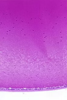
Lens of the eye. Light micrograph of a section through the lens from a human eye. The lens consists of thousands of precisely aligned stacks of fibres seen here end-on

Background imageTissue Collection: Heart tissue, SEM C015 / 9541

Heart tissue, SEM C015 / 9541
Heart tissue. Coloured scanning electron micrograph (SEM) of heart tissue with red blood cells (erythrocytes)

Background imageTissue Collection: Heart muscle, light micrograph C016 / 0517

Heart muscle, light micrograph C016 / 0517
Heart muscle. Light micrograph of a section through heart (cardiac) muscle tissue, showing cardiomyocyte muscle cells. These cells have a centrally located nucleus and are branched

Background imageTissue Collection: Brain glial cells, light micrograph C016 / 0524

Brain glial cells, light micrograph C016 / 0524
Brain glial cells. Light micrograph of a section through brain tissue, showing numerous glial cells (dark). Glial cells, or neuroglia, are non-neural cells of the central nervous system (CNS)

Background imageTissue Collection: Cirrhosis of liver, light micrograph C016 / 0530

Cirrhosis of liver, light micrograph C016 / 0530
Cirrhosis of liver. Light micrograph of a section through liver tissue, damaged by cirrhosis. Cirrhosis occurs as a result of a range of factors causing damage to liver function

Background imageTissue Collection: Liver, SEM C015 / 9923

Liver, SEM C015 / 9923
Liver vein. Coloured scanning electron micrograph (SEM) of liver tissue. The functions of the liver include destroying old red blood cells

Background imageTissue Collection: Pancreas lobes, SEM C015 / 9495

Pancreas lobes, SEM C015 / 9495
Pancreas lobes, coloured scanning electron micrograph (SEM). Several lobes of the pancreas are seen here, separated by fissures. The smaller sections seen on each lobe are clusters of acini cells

Background imageTissue Collection: Stratified squamous epithelium C016 / 0534

Stratified squamous epithelium C016 / 0534
Stratified squamous epithelium. Light micrograph of a section through stratified squamous epithelium, showing the multiple layers, or strata, that function to resist abrasion of the surface

Background imageTissue Collection: Nerve ganglion, light micrograph C016 / 0532

Nerve ganglion, light micrograph C016 / 0532
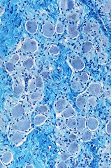
Nerve ganglion. Light micrograph of a section through a nerve ganglion of the peripheral nervous system, showing clusters of nerve cell bodies

Background imageTissue Collection: Myelinated nerve fibres, light micrograph C016 / 0531

Myelinated nerve fibres, light micrograph C016 / 0531
Myelinated nerve fibres. Light micrograph of a section through a spinal sensory nerve containing many closely packed axons exhibiting a wave-type appearance

Background imageTissue Collection: Pancreas cells, SEM C015 / 9503

Pancreas cells, SEM C015 / 9503
Pancreas cells, coloured scanning electron micrograph (SEM). The small sections seen here are clusters of acini cells. These are exocrine cells, secreting digestive enzymes


All Professionally Made to Order for Quick Shipping

"Tissue: A Microscopic World Unveiled" Uterus lining during menstruation, SEM: Witness the intricate dance of life as the uterus sheds its lining during menstruation, revealing the resilience and beauty within. Dicotyledon plant stem, light micrograph: Delve into the mesmerizing patterns etched on a dicotyledon plant stem under a microscope's gaze, showcasing nature's artistry in every fiber. Skin disorders, artwork: Explore an artistic interpretation of skin disorders that unveils the hidden struggles faced by individuals while highlighting their strength and resilience. Brain tissue blood supply: Uncover the lifeline of our thoughts and emotions as we delve into the intricate network of blood vessels supplying oxygen to brain tissues - a testament to our cognitive prowess. Botanik Digitalis purpurea L. Fingerhut 160: 1: Immerse yourself in the captivating world of Botanik Digitalis purpurea L. , where delicate petals intertwine with vibrant hues, offering solace amidst nature's grandeur. Lactating breast tissue, light micrograph: Marvel at motherhood's miracle through a gentle lens capturing lactating breast tissue – an exquisite portrayal of nourishment and love personified. Blood cells: Embark on a journey within our veins as we explore blood cells bustling with life; each cell holds secrets that unlock vital clues about our health and well-being. Pine pollen grains, light micrograph: Peer into nature's microscopic marvels as pine pollen grains come alive under magnification – tiny particles carrying life across vast distances in pursuit of pollination perfection. Pine stem, light micrograph: Discover the sturdy elegance concealed beneath rough bark as you witness a pine stem reveal its inner intricacies through delicate beams of light – strength rooted deep within nature itself. Lime tree stem, light micrograph.