Fraud Blocker Skip to main content

Tissue Collection (page 4)

"Tissue: A Microscopic World Unveiled" Uterus lining during menstruation, SEM: Witness the intricate dance of life as the uterus sheds its lining during menstruation

Background imageTissue Collection: Retina layers, SEM C018 / 0520

Retina layers, SEM C018 / 0520
Retina layers, coloured scanning electron micrograph (SEM). The retina of the human eye is found on the inside of the eyeball and is formed from a number of layers
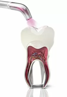
Background imageTissue Collection: Dental filling polymerisation, artwork C014 / 2025

Dental filling polymerisation, artwork C014 / 2025
Dental filling polymerisation. Artwork of a cross-section of a human tooth, with a filling (white) being set using an ultraviolet light lamp tool

Background imageTissue Collection: Human olfactory system, artwork C013 / 5940

Human olfactory system, artwork C013 / 5940
Human olfactory system. Cutaway computer artwork of a persons head, with an enlargement (upper right) showing the olfactory receptor cells (red)

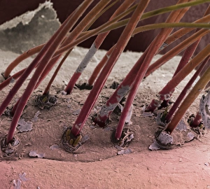
Background imageTissue Collection: Eyelash hairs, SEM

Eyelash hairs, SEM
Eyelash hairs and skin. Coloured scanning electron micrograph (SEM) of eyelash hairs growing from the surface of human skin

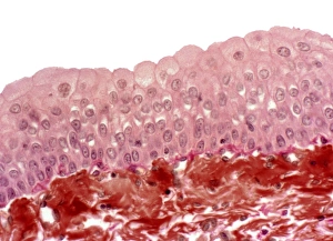
Background imageTissue Collection: Bladder epithelium, light micrograph

Bladder epithelium, light micrograph
Bladder epithelium. Light micrograph of a vertical section through the wall of the urinary bladder. The inner surface is at top

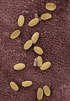
Background imageTissue Collection: Allergens in trachea

Allergens in trachea. Coloured scanning electron micrograph (SEM) of the surface of the trachea (windpipe) with a number of pollen grains (yellow) attached

Background imageTissue Collection: Trachea, SEM

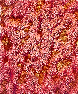
Trachea, SEM
Trachea. Coloured scanning electron micrograph (SEM) of the lining of the trachea (windpipe). The trachea links the larynx (voice box) to the lungs

Background imageTissue Collection: Osteoblast bone cell, SEM

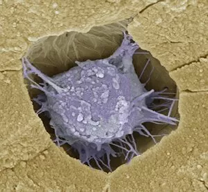
Osteoblast bone cell, SEM
Osteoblast bone cell. Coloured scanning electron micrograph (SEM) of an osteoblast (centre) in bone tissue. Osteoblasts are primarily found in regions of new bone growth

Background imageTissue Collection: Whale bone tissue, light micrograph

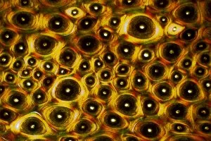
Whale bone tissue, light micrograph

Background imageTissue Collection: Hazel nut, light micrograph

Hazel nut, light micrograph
Hazel nut. Light micrograph of a section through a nut from a common hazel (Corylus avellana) tree, showing the large amount of fat (red) stored within endosperm tissue

Background imageTissue Collection: Skin and hair follicles, light micrograph

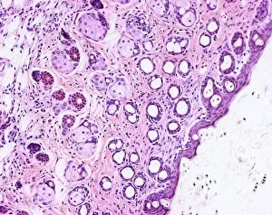
Skin and hair follicles, light micrograph
Skin and hair follicles. Light micrograph of a section through skin, showing hair follicles (dark purple circular structures). Haematoxylin and eosin stain

Background imageTissue Collection: Iris root, light micrograph

Iris root, light micrograph
Iris root. Light micrograph of a section through the root of an Iris plant (Iris germanica) showing a vascular cylinder. The cylinder is comprised of a central cluster of parenchyma cells (red)

Background imageTissue Collection: Quiver tree and moon in early morning light Augrabis Falls National Park, Northern Cape Province

Quiver tree and moon in early morning light Augrabis Falls National Park, Northern Cape Province, South Africa
SAS-325 Kokerboom / Quiver / Aloe Trees - and moon - in early morning light Augrabis Falls National Park, Northern Cape Province

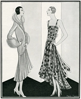
Background imageTissue Collection: Evening gown and fur coat

Evening gown and fur coat
An evening dress with trains that only just touch the ground expressed in tissue lame from Vanit霧s of Sloane Street, paired with a mink and musquash coat from Artemis. 1929

Background imageTissue Collection: Evening dress and coatee from Peter Robinson

Evening dress and coatee from Peter Robinson
A dress and coat of metal lace mounted on tissue, the latter trimmed with fur. A telling touch of colour is introduced by the velvet loop and pendant ends caught with a crystal ornament

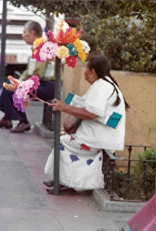
Background imageTissue Collection: Mexican woman sitting with paper tissue flower arrangments

Mexican woman sitting with paper tissue flower arrangments. Dressed in white with two long black plaits. A man sits further down the picture, in Tijuana Mexico. Date: 1982

Background imageTissue Collection: Street art festival in Tijuana Mexico

Street art festival in Tijuana Mexico
1982 Street art festival in Tijuana Mexico. Mexican men and women carrying paper tissue flower arrangements on the crowed street. Date: 1982

Background imageTissue Collection: Torah mantle. Sefrou, Morroco, ca. 1926. Israel Museum. Jeru

Torah mantle. Sefrou, Morroco, ca. 1926. Israel Museum. Jeru
Torah mantle. Sefrou, Morroco, ca. 1926. Cotton, silk; abd gilt metal-thread embroidery. Inscribed in Hebrew. Israel Museum. Jerusalem

Background imageTissue Collection: Fashions for the London Season by Gordon Conway

Fashions for the London Season by Gordon Conway
Nothing could be smarter than this evening coat and dress from Dickins and Jones, Regent Street. The former is of gold-tinsel tissue shot with moonlight blue, this gives a peach-bloom effect

Background imageTissue Collection: Islamic period. Spain. Fragment layer. Silk fabric. 11th C

Islamic period. Spain. Fragment layer. Silk fabric. 11th C. From Santa Maria de la Seu D Urgell. Diocesan Museum. Seu D Urgell. Catalonia. Spain

Background imageTissue Collection: Tissue fragments. 13th century. Diocesan Museum. La Seu d Ur

Tissue fragments. 13th century. Diocesan Museum. La Seu d Urgell. Catalonia. Spain

Background imageTissue Collection: Tissue fragments. 12th-13th centuries. Diocesan Museum. La S

Tissue fragments. 12th-13th centuries. Diocesan Museum. La Seu d Urgell. Catalonia. Spain

Background imageTissue Collection: Islamic art. Spain. 12th-13th c. Tissue of La Seu d Urgell

Islamic art. Spain. 12th-13th c. Tissue of La Seu d Urgell. Episcopal Museum of Vic. Catalonia

Background imageTissue Collection: Hispano-Arabic. Piece of tissue with eagles. 12th-13th c. Li

Hispano-Arabic. Piece of tissue with eagles. 12th-13th c. Li
Textile. Hispano-Arabic. Piece of tissue with eagles. 12th-13th century. Linen and silk. From Collegiate of St. Vicenc de Roda de Ribagorca. Episcopal Museum of Vic. Catalonia. Spain

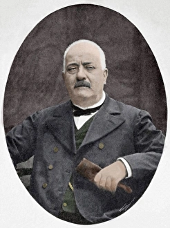
Background imageTissue Collection: Ramon Batlle Ribas (1837-1906). Photography

Ramon Batlle Ribas (1837-1906). Photography
Ramon Batlle Ribas (1837-1906). Spanish technical and textile teacher. Founder of the first school for teaching mechanical tissue

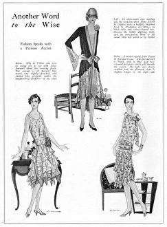
Background imageTissue Collection: Three Parisian fashion models, 1927

Three Parisian fashion models, 1927, from Alice Bernard (evening frock), Madeleine des Hayes (race frock) and Patou (flowered tissue frock) 1927

Background imageTissue Collection: Suffrage Demonstration N. U. W. S.s 1908

Suffrage Demonstration N. U. W. S.s 1908
A souvenir of the Womens Suffrage March and Mass Meeting at the Albert Hall, Saturday, June 13, 1908. Gives details of the march from Embankment to the Albert Hall

Background imageTissue Collection: Tissue Manufacture

Tissue Manufacture
A group of Russian women whiten tissues in a field. Date: circa 1900

Background imageTissue Collection: Two Women Sewing Lamplight ca 1853 Zinc plate etching

Two Women Sewing Lamplight ca 1853 Zinc plate etching
Artokoloro

Background imageTissue Collection: Twenty-Three Sheets Drawings Glassware Mirrors

Twenty-Three Sheets Drawings Glassware Mirrors
Artokoloro

Background imageTissue Collection: Design nave ca 1892-1918 New York United States

Design nave ca 1892-1918 New York United States
Artokoloro

Background imageTissue Collection: Arthur Haden 1859 Drypoint fourth state four

Arthur Haden 1859 Drypoint fourth state four
Artokoloro

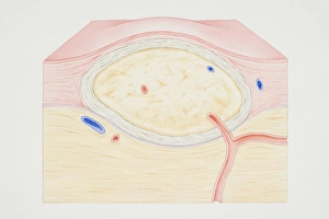
Background imageTissue Collection: Cross-section diagram of a non-cancerous tumour including a fibrous capsule

Cross-section diagram of a non-cancerous tumour including a fibrous capsule, tissue layer and blood vessel

Background imageTissue Collection: Breast showing rib, pectoral muscle, lung, fatty tissue, blood vessel, lobule, ampulla, nipple

Breast showing rib, pectoral muscle, lung, fatty tissue, blood vessel, lobule, ampulla, nipple, areola and milk duct, cross-section

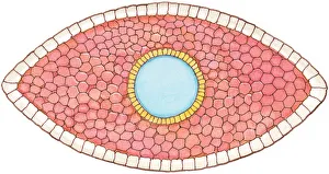
Background imageTissue Collection: Illustration of cross section showing how early stage embryo of Flatworm (Phylum Platyhelminthes)

Illustration of cross section showing how early stage embryo of Flatworm (Phylum Platyhelminthes) differentiates into three layers of cell tissue, ectoderm, mesoderm and endoderm

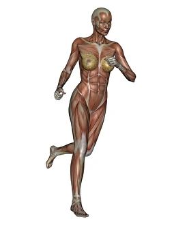
Background imageTissue Collection: Muscular woman running

Muscular woman running, isolated on white background

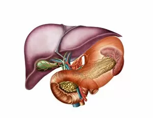
Background imageTissue Collection: Anatomy of liver, antero-visceral view

Anatomy of liver, antero-visceral view

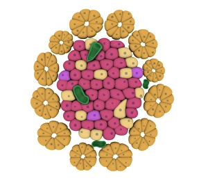
Background imageTissue Collection: Conceptual image of pancreatic islet of Langerhans

Conceptual image of pancreatic islet of Langerhans

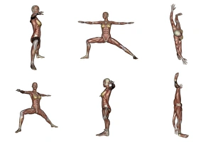
Background imageTissue Collection: Six different views of warrior yoga pose showing female musculature

Six different views of warrior yoga pose showing female musculature, white background

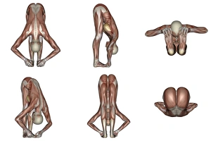
Background imageTissue Collection: Six different views of big toes yoga pose showing female musculature

Six different views of big toes yoga pose showing female musculature, white background

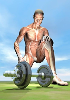
Background imageTissue Collection: Male musculature looking at a dumbbell on the grass

Male musculature looking at a dumbbell on the grass

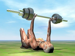
Background imageTissue Collection: Female musculature exercising with a dumbbell

Female musculature exercising with a dumbbell while lying down on the grass

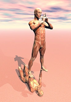
Background imageTissue Collection: Female lying on the ground holding a male upon her two hands

Female lying on the ground holding a male upon her two hands
Female lying on the ground and holding a peaceful male upon her two hands, musculature view

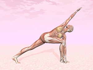
Background imageTissue Collection: Female musculature performing revolved side angle yoga pose

Female musculature performing revolved side angle yoga pose, pink background

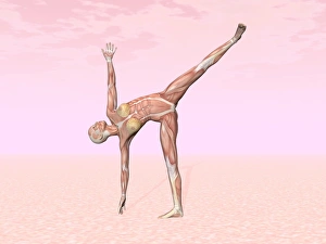
Background imageTissue Collection: Female musculature performing half moon yoga pose

Female musculature performing half moon yoga pose, pink background

Background imageTissue Collection: Osteoclast breaking down bone tissue

Osteoclast breaking down bone tissue

Background imageTissue Collection: Two male musculatures fighting martial arts

Two male musculatures fighting martial arts

Background imageTissue Collection: Microscopic view of Anthrax

Microscopic view of Anthrax. Anthrax is an acute disease caused by the bacterium Bacillus anthracis. Most forms of the disease are lethal, and it affects both humans and animals


All Professionally Made to Order for Quick Shipping

"Tissue: A Microscopic World Unveiled" Uterus lining during menstruation, SEM: Witness the intricate dance of life as the uterus sheds its lining during menstruation, revealing the resilience and beauty within. Dicotyledon plant stem, light micrograph: Delve into the mesmerizing patterns etched on a dicotyledon plant stem under a microscope's gaze, showcasing nature's artistry in every fiber. Skin disorders, artwork: Explore an artistic interpretation of skin disorders that unveils the hidden struggles faced by individuals while highlighting their strength and resilience. Brain tissue blood supply: Uncover the lifeline of our thoughts and emotions as we delve into the intricate network of blood vessels supplying oxygen to brain tissues - a testament to our cognitive prowess. Botanik Digitalis purpurea L. Fingerhut 160: 1: Immerse yourself in the captivating world of Botanik Digitalis purpurea L. , where delicate petals intertwine with vibrant hues, offering solace amidst nature's grandeur. Lactating breast tissue, light micrograph: Marvel at motherhood's miracle through a gentle lens capturing lactating breast tissue – an exquisite portrayal of nourishment and love personified. Blood cells: Embark on a journey within our veins as we explore blood cells bustling with life; each cell holds secrets that unlock vital clues about our health and well-being. Pine pollen grains, light micrograph: Peer into nature's microscopic marvels as pine pollen grains come alive under magnification – tiny particles carrying life across vast distances in pursuit of pollination perfection. Pine stem, light micrograph: Discover the sturdy elegance concealed beneath rough bark as you witness a pine stem reveal its inner intricacies through delicate beams of light – strength rooted deep within nature itself. Lime tree stem, light micrograph.