Fraud Blocker Skip to main content

Tissue Collection (page 7)

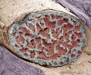
"Tissue: A Microscopic World Unveiled" Uterus lining during menstruation, SEM: Witness the intricate dance of life as the uterus sheds its lining during menstruation

Background imageTissue Collection: Woman with a cold F005 / 0307

Woman with a cold F005 / 0307
MODEL RELEASED. Woman with a cold

Background imageTissue Collection: Plant surface, artwork

Plant surface, artwork
Plant surface, computer artwork

Background imageTissue Collection: Kidney cancer, light micrograph F005 / 6071

Kidney cancer, light micrograph F005 / 6071
Kidney cancer. Light micrograph of a section through a Wilms tumour of the kidney. This solid tumour generally affects children, with most cases occurring between 3 and 4 years of age

Background imageTissue Collection: Human skin anatomy, artwork

Human skin anatomy, artwork
Human skin anatomy, computer artwork

Background imageTissue Collection: Vocal cords, artwork

Vocal cords, artwork
Vocal cords. Computer artwork of a rear view of the larynx (purple/grey), also known as the voice box, and the vocal cords (centre)

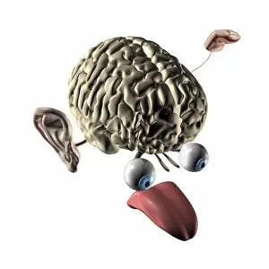
Background imageTissue Collection: Human brain and senses, artwork

Human brain and senses, artwork
Human brain and senses. Artwork of a human brain, seen obliquely from the front, with the five senses shown. These are the visual organs (vision, the eyes), the olfactory organ (smell, the nose)

Background imageTissue Collection: Small intestine, light micrograph

Small intestine, light micrograph
Small intestine. Light micrograph of a section through the small intestine. The finger-like projections at left are the villi. These increase the surface area for the absorption of food

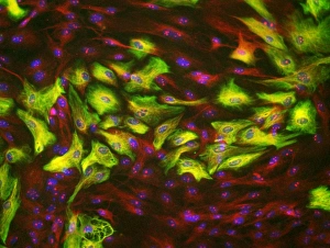
Background imageTissue Collection: Astrocyte brain cells, light micrograph

Astrocyte brain cells, light micrograph
Astrocyte brain cells. Fluorescence light micrograph of primary astrocyte cells from the brain of a mouse. Astrocytes have numerous branches of connective tissue that provide support

Background imageTissue Collection: Hair follicle, SEM C014 / 0382

Hair follicle, SEM C014 / 0382
Hair follicle. Coloured scanning electron micrograph (SEM) of a section through a hair shaft and follicle (round structures at centre)

Background imageTissue Collection: Woman blowing her nose F006 / 2637

Woman blowing her nose F006 / 2637
MODEL RELEASED. Woman blowing her nose

Background imageTissue Collection: Woman blowing her nose F006 / 2636

Woman blowing her nose F006 / 2636
MODEL RELEASED. Woman blowing her nose

Background imageTissue Collection: Woman blowing her nose F006 / 2635

Woman blowing her nose F006 / 2635
MODEL RELEASED. Woman blowing her nose

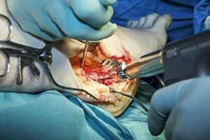
Background imageTissue Collection: Ankle surgery: dissecting out tissues C014 / 7781

Ankle surgery: dissecting out tissues C014 / 7781
Ankle surgery. Close-up of surgical tools being used to dissect tissue from a patients ankle. The tool at right is a bone saw, with retractors being used to keep the surgical incision open

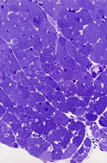
Background imageTissue Collection: Lactating breast tissue, light micrograph F006 / 9797

Lactating breast tissue, light micrograph F006 / 9797
Lactating breast tissue, light micrograph. Magnification: x150 when printed at 10 centimetres wide
Background imageTissue Collection: Hair follicle, SEM C014 / 0383

Hair follicle, SEM C014 / 0383
Hair follicle. Coloured scanning electron micrograph (SEM) of a section through a hair shaft and follicle (round structures at centre)

Background imageTissue Collection: Surgical Instruments. C017 / 3470

Surgical Instruments. C017 / 3470
Plate from 18th century encyclopedia showing illustrations of various surgical instruments. A surgical instrument is a specially designed tool or device for performing specific actions such as

Background imageTissue Collection: Brain nerve cells, TEM C014 / 0357

Brain nerve cells, TEM C014 / 0357
Brain nerve cells. Transmission electron micrograph (TEM) of a section through brain tissue from the cerebral cortex, showing numerous neurons (nerve cells) surrounded by axons and dendrites

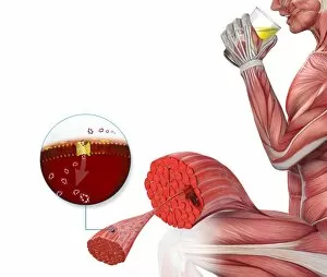
Background imageTissue Collection: Sugar uptake in muscles, diagram

Sugar uptake in muscles, diagram
Sugar uptake in muscles. Diagram showing sugar from a drink (upper right) being taken up by the muscles of the human body

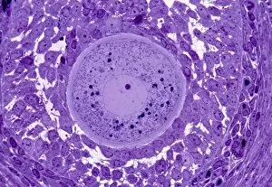
Background imageTissue Collection: Oocyte, light micrograph

Oocyte, light micrograph
Oocyte. Light micrograph of a section through an oocyte within a early antral follicle in an ovary. Oocytes are immature ova, or egg cells

Background imageTissue Collection: Cardiac muscle, light micrograph

Cardiac muscle, light micrograph
Cardiac muscle. Light micrograph of a section through muscle tissue from a heart, showing the cardiomyocyte cells. Heart muscle cells show central nuclei but the dominant feature is the abundance of

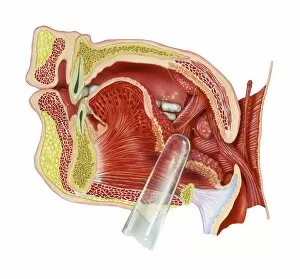
Background imageTissue Collection: Tonsils, artwork

Tonsils, artwork
Tonsils. Artwork of a section through the mouth showing a palatine tonsil. The clear rod points to the tonsillar fossa, a depression that is occupied by a palatine tonsil (round, right)

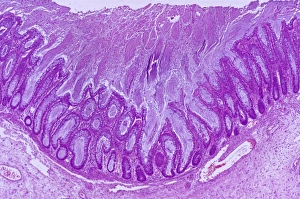
Background imageTissue Collection: Colitis, light micrograph

Colitis, light micrograph
Colitis. Light micrograph of a section through colon (large intestine) tissue affected by bacterial infection, causing injury to the mucosal lining, which has resulted in inflammation (colitis)

Background imageTissue Collection: Hairy scalp skin, light micrograph

Hairy scalp skin, light micrograph
Hairy scalp skin. Light micrograph of a section through healthy skin from a human scalp, showing the numerous hair follicles. The scalp is an example of hairy skin

Background imageTissue Collection: Oxalosis, light micrograph

Oxalosis, light micrograph
Oxalosis. Light micrograph of a section through adult bone tissue affected by oxalosis (primary hyperoxaluria), showing mature bone (green)

Background imageTissue Collection: Peripheral nerve, light micrograph

Peripheral nerve, light micrograph
Peripheral nerve. Light micrograph of a section through a peripheral nerve. This is a mixed nerve with myelinated axons (dark blue circles)

Background imageTissue Collection: Ischaemic bowel, light micrograph

Ischaemic bowel, light micrograph
Ischaemic bowel. Light micrograph of a section through a portion of intestine that has been damaged by an interruption of the blood supply, or ischaemia

Background imageTissue Collection: Nerve ganglion, light micrograph

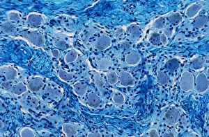
Nerve ganglion, light micrograph
Nerve ganglion. Light micrograph of a section through a dorsal (sensory) spinal root ganglion associated with a sensory nerve root of the spinal cord. Sensory information from peripheral sites e.g

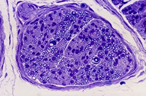
Background imageTissue Collection: Myelinated nerve, light micrograph

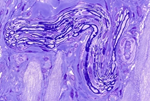
Myelinated nerve, light micrograph
Myelinated nerve. Light micrograph of a section through a peripheral myelinated nerve, showing many individual axons each covered with deep blue-staining myelin sheaths

Background imageTissue Collection: Glial cells, light micrograph

Glial cells, light micrograph
Glial cells in the brain. Light micrograph of a section through glial cells (dark) in the grey matter of the brain. Due to their star shape these glial cells are called astrocytes

Background imageTissue Collection: Fat tissue, artwork

Fat tissue, artwork
Block of cells in adipose tissue

Background imageTissue Collection: Facial anatomy, artwork

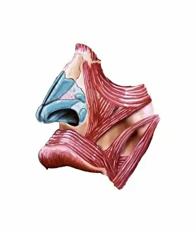
Facial anatomy, artwork
Facial anatomy. Computer artwork showing the superficial muscles around the nose and upper lip of a human face

Background imageTissue Collection: Fibrolipoma of the spine, MRI scan

Fibrolipoma of the spine, MRI scan
Fibrolipoma of the spine. Coloured magnetic resonance imaging (MRI) scan of a sagittal section through the thoraco-lumbar spine of a 19-month-old patient

Background imageTissue Collection: Cirrhosis of the liver, light micrograph

Cirrhosis of the liver, light micrograph
Cirrhosis of the liver. Light micrograph of a section through liver tissue affected by cirrhosis, showing a cluster of hepatocyte cells (orange)

Background imageTissue Collection: Lassa virus particles, TEM C016 / 9409

Lassa virus particles, TEM C016 / 9409
Lassa virus particles. Transmission electron micrograph (TEM) of Lassa virus particles (virions, blue) amongst cell debris. This Arenavirus is the cause of Lassa fever

Background imageTissue Collection: Fimbriae of a fallopian tube, SEM C016 / 9047

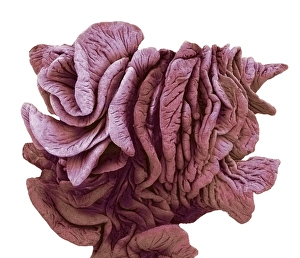
Fimbriae of a fallopian tube, SEM C016 / 9047
Fimbriae of a fallopian tube, coloured scanning electron micrograph (SEM). Fimbriae are the folds at the opening (ostium) of a fallopian tube, next to an ovary

Background imageTissue Collection: SARS virus particles, TEM C016 / 9445

SARS virus particles, TEM C016 / 9445
SARS virus particles. Coloured transmission electron micrograph (TEM) of a section through a tissue sample infected with numerous SARS coronavirus particles (virions, dark, round)

Background imageTissue Collection: Acute haemorrhagic conjunctivitis, TEM C016 / 9387

Acute haemorrhagic conjunctivitis, TEM C016 / 9387
Acute haemorrhagic conjunctivitis. Transmission electron micrograph (TEM) of a sample of tissue from the conjunctiva of a human eye that is infected by enterovirus particles (purple)

Background imageTissue Collection: Eastern equine encephalitis, TEM C016 / 9390

Eastern equine encephalitis, TEM C016 / 9390
Eastern equine encephalitis. Transmission electron micrograph (TEM) of a section through tissue from a human central nervous system (CNS)

Background imageTissue Collection: Colorado tick fever virus infection, TEM C016 / 9384

Colorado tick fever virus infection, TEM C016 / 9384
Colorado tick fever virus infection. Transmission electron micrograph (TEM) of human tissue infected by Colorado tick fever virus (CTFV) particles (virions, dark circles)

Background imageTissue Collection: Broken finger bone, SEM C016 / 9027

Broken finger bone, SEM C016 / 9027
Broken finger bone. Coloured scanning electron micrograph (SEM) showing the internal structure of a broken finger bone (phalanx)

Background imageTissue Collection: Swine flu virus particles, TEM C016 / 9399

Swine flu virus particles, TEM C016 / 9399
Swine flu virus particles. Transmission electron micrograph (TEM) of a section through H3N2 influenza A virus particles (virions)

Background imageTissue Collection: Swine flu virus particles, TEM C016 / 9406

Swine flu virus particles, TEM C016 / 9406
Influenza virus particles. Transmission electron micrograph (TEM) of influenza (flu) virus particles (virions). Each virion consists of ribonucleic acid (RNA, dark patches)

Background imageTissue Collection: Dicotyledon root, SEM C016 / 9125

Dicotyledon root, SEM C016 / 9125
Dicotyledon root. Coloured scanning electron micrograph (SEM) of a transverse section through an immature root from a dicotyledonous plant

Background imageTissue Collection: St. Louis encephalitis virus particles C016 / 9453

St. Louis encephalitis virus particles C016 / 9453
St. Louis encephalitis virus particles. Coloured transmission electron micrograph (TEM) of a section through a mosquito salivary gland containing numerous Saint Louis encephalitis virus particles

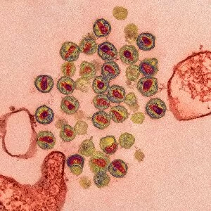
Background imageTissue Collection: HIV virus particles, TEM C016 / 9404

HIV virus particles, TEM C016 / 9404
HIV virus particles. Transmission electron micrograph (TEM) of a section through human immunodeficiency virus (HIV) particles (virions, round)

Background imageTissue Collection: Smallpox virus particles, TEM C016 / 9447

Smallpox virus particles, TEM C016 / 9447
Smallpox virus particles. Coloured transmission electron micrograph (TEM) of a section through a tissue sample infected with numerous Variola virus particles (virions, brown)

Background imageTissue Collection: Dicotyledon vascular bundle, SEM C016 / 9603

Dicotyledon vascular bundle, SEM C016 / 9603
Dicotyledon vascular bundle. Coloured scanning electron micrograph (SEM) of a section through a vascular bundle from a dicotyledonous plant

Background imageTissue Collection: Spumavirus particles, TEM C016 / 9452

Spumavirus particles, TEM C016 / 9452
Spumavirus virus particles. Coloured transmission electron micrograph (TEM) of a section through a tissue sample infected with numerous spumavirus particles (virions, yellow)


All Professionally Made to Order for Quick Shipping

"Tissue: A Microscopic World Unveiled" Uterus lining during menstruation, SEM: Witness the intricate dance of life as the uterus sheds its lining during menstruation, revealing the resilience and beauty within. Dicotyledon plant stem, light micrograph: Delve into the mesmerizing patterns etched on a dicotyledon plant stem under a microscope's gaze, showcasing nature's artistry in every fiber. Skin disorders, artwork: Explore an artistic interpretation of skin disorders that unveils the hidden struggles faced by individuals while highlighting their strength and resilience. Brain tissue blood supply: Uncover the lifeline of our thoughts and emotions as we delve into the intricate network of blood vessels supplying oxygen to brain tissues - a testament to our cognitive prowess. Botanik Digitalis purpurea L. Fingerhut 160: 1: Immerse yourself in the captivating world of Botanik Digitalis purpurea L. , where delicate petals intertwine with vibrant hues, offering solace amidst nature's grandeur. Lactating breast tissue, light micrograph: Marvel at motherhood's miracle through a gentle lens capturing lactating breast tissue – an exquisite portrayal of nourishment and love personified. Blood cells: Embark on a journey within our veins as we explore blood cells bustling with life; each cell holds secrets that unlock vital clues about our health and well-being. Pine pollen grains, light micrograph: Peer into nature's microscopic marvels as pine pollen grains come alive under magnification – tiny particles carrying life across vast distances in pursuit of pollination perfection. Pine stem, light micrograph: Discover the sturdy elegance concealed beneath rough bark as you witness a pine stem reveal its inner intricacies through delicate beams of light – strength rooted deep within nature itself. Lime tree stem, light micrograph.