Fraud Blocker Skip to main content

Proteomics Collection (page 4)

Proteomics, the study of proteins and their functions within an organism, is a fascinating field that unravels the intricate workings of life

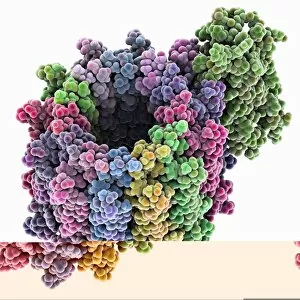
Background imageProteomics Collection: Potassium ion channel cavity structure

Potassium ion channel cavity structure. Molecular model showing the structure of a cavity formed by potassium ion channel proteins
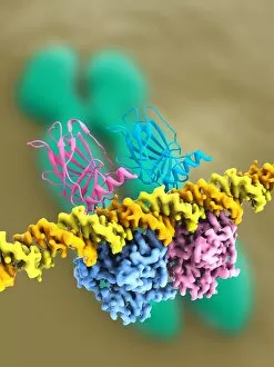
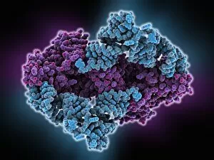
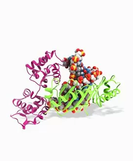
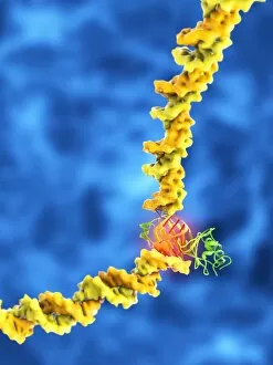
Background imageProteomics Collection: Tumour suppressor protein and DNA C017 / 3645

Tumour suppressor protein and DNA C017 / 3645
Tumour suppressor protein and DNA. Computer artwork showing a molecule of the tumour suppressor protein p53 (blue and pink) bound to a molecule of DNA (deoxyribonucleic acid, yellow and orange)

Background imageProteomics Collection: ATP synthase molecule C014 / 0880

ATP synthase molecule C014 / 0880
ATP synthase molecule. Molecular model showing the structure of ATP synthase (ATPase) subunit C. ATPase is an important enzyme that provides energy for cells through the synthesis of adenosine

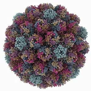
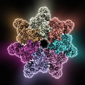
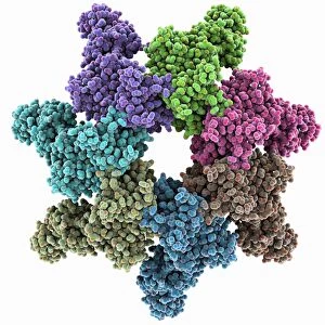
Background imageProteomics Collection: Avian polyomavirus capsid

Avian polyomavirus capsid, molecular model. This virus, one of a range named for their potential to cause multiple tumours, infects birds. Discovered in budgerigars in 1981, it is often fatal

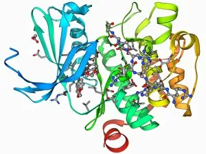
Background imageProteomics Collection: Anthrax protective antigen molecule C014 / 0886

Anthrax protective antigen molecule C014 / 0886
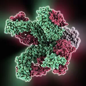
Anthrax protective antigen molecule. Computer model showing the structure of a molecule of protective antigen (PA) produced by anthrax (Bacillus anthracis) bacteria

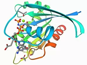
Background imageProteomics Collection: HIV enzyme protein, molecular model C014 / 0876

HIV enzyme protein, molecular model C014 / 0876
HIV enzyme protein. Computer model showing the structure of the catalytic domain of a molecule of HIV-1 retroviral integrase (IN) from the human immunodeficiency virus (HIV)

Background imageProteomics Collection: Tryptophanyl-tRNA synthetase molecule

Tryptophanyl-tRNA synthetase molecule
Tryptophanyl-tRNA synthetase protein molecule. Molecular model showing human tryptophanyl-tRNA synthetase complexed with tryptophan tRNA (transfer ribonucleic acid)

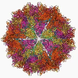
Background imageProteomics Collection: Cytoplasmic polyhedrosis virus capsid

Cytoplasmic polyhedrosis virus capsid, molecular model. Part of the Cypovirus genus and invariably fatal, this insect virus is transmitted by contamination of leaves eaten (examples include silkworms)

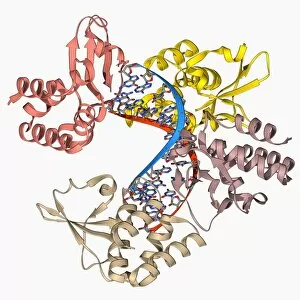
Background imageProteomics Collection: Pho4 transcription factor bound to DNA C014 / 0861

Pho4 transcription factor bound to DNA C014 / 0861
Pho4 transcription factor bound to DNA. Molecular model showing phosphate system positive regulatory protein (Pho4) (blue and green) bound to a strand of DNA (deoxyribonucleic acid, red and purple)

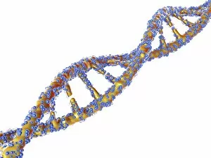
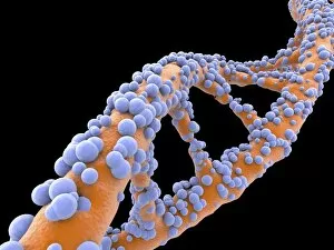
Background imageProteomics Collection: DNA molecule, artwork F007 / 1996

DNA molecule, artwork F007 / 1996
DNA molecule, computer artwork

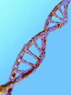
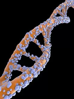
Background imageProteomics Collection: DNA molecule, artwork F007 / 1994

DNA molecule, artwork F007 / 1994
DNA molecule, computer artwork

Background imageProteomics Collection: DNA molecule, artwork F007 / 1995

DNA molecule, artwork F007 / 1995
DNA molecule, computer artwork

Background imageProteomics Collection: DNA molecule, artwork F007 / 1991

DNA molecule, artwork F007 / 1991
DNA molecule, computer artwork

Background imageProteomics Collection: DNA molecule, artwork F007 / 1992

DNA molecule, artwork F007 / 1992
DNA molecule, computer artwork

Background imageProteomics Collection: HIV enzyme protein, molecular model

HIV enzyme protein, molecular model
HIV enzyme protein. Computer model showing the structure of the catalytic domain of a molecule of HIV-1 retroviral integrase (IN) from the human immunodeficiency virus (HIV)

Background imageProteomics Collection: Anthrax protective antigen molecule C014 / 0865

Anthrax protective antigen molecule C014 / 0865
Anthrax protective antigen molecule. Computer model showing the structure of a molecule of protective antigen (PA) produced by anthrax (Bacillus anthracis) bacteria

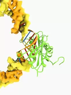
Background imageProteomics Collection: TATA box-binding protein complex C017 / 7090

TATA box-binding protein complex C017 / 7090
TATA box-binding protein complex. Molecular model showing a TATA box-binding protein (TBP, green) complexed with a strand of DNA (deoxyribonucleic acid, spheres) and transcription factor IIB

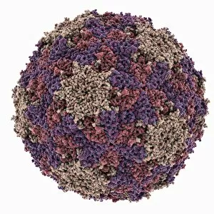
Background imageProteomics Collection: Theilers encephalomyelitis virus capsid

Theilers encephalomyelitis virus capsid, molecular model. This virus, which causes brain and spinal cord inflammation in mice, is used in research

Background imageProteomics Collection: TATA box-binding protein complex C017 / 7085

TATA box-binding protein complex C017 / 7085
TATA box-binding protein complex. Molecular model showing a TATA box-binding protein (TBP, green) complexed with a strand of DNA (deoxyribonucleic acid, yellow) and transcription factor IIB

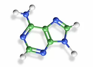
Background imageProteomics Collection: Adenine molecule, artwork C017 / 7199

Adenine molecule, artwork C017 / 7199
Adenine molecule. Computer artwork showing the structure of a molecule of the nucleobase adenine. Atoms are colour-coded spheres: carbon (green), nitrogen (blue), and oxygen (white)

Background imageProteomics Collection: Ricin A-chain, artwork C017 / 3654

Ricin A-chain, artwork C017 / 3654
Ricin A-chain. Computer artwork showing the enzymatically active A-chain from a molecule of the toxic protein ricin. Ricin comprises two entwined amino acid chains; A (seen here) and B (not shown)

Background imageProteomics Collection: Sirtuin enzyme and p53, artwork C017 / 3660

Sirtuin enzyme and p53, artwork C017 / 3660
Sirtuin enzyme and p53. Computer artwork of a sirtuin (Sir2) enzyme (blue) bound to a p53 peptide (pink). Sir2 enzymes form a unique class of NAD(+)

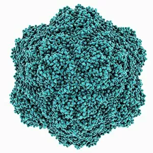
Background imageProteomics Collection: Tobacco necrosis virus capsid

Tobacco necrosis virus capsid, molecular model. This plant virus infects a wide rage of plants, including the tobacco plant for which it is named. The virus causes tissue death (necrosis)

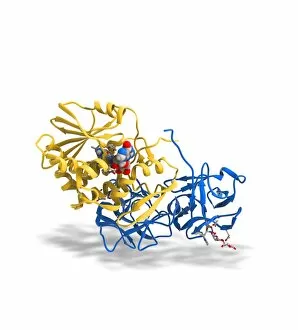
Background imageProteomics Collection: Ricin molecule, artwork C017 / 3649

Ricin molecule, artwork C017 / 3649
Ricin molecule. Computer artwork showing the structure of a molecule of the toxic protein ricin. Ricin comprises two entwined amino acid chains; A (yellow) and B (blue)

Background imageProteomics Collection: TATA box-binding protein complex C017 / 7083

TATA box-binding protein complex C017 / 7083
TATA box-binding protein complex. Molecular model showing a TATA box-binding protein (TBP, green) complexed with a strand of DNA (deoxyribonucleic acid, yellow) and transcription factor IIB

Background imageProteomics Collection: Rubisco enzyme molecule F006 / 9776

Rubisco enzyme molecule F006 / 9776
Rubisco. Molecular model of the enzyme rubisco (ribulose bisphosphate carboxylase oxygenase) complexed with 2-carboxyarabinitol biphosphate

Background imageProteomics Collection: Rubisco enzyme molecule F006 / 9779

Rubisco enzyme molecule F006 / 9779
Rubisco. Molecular model of the enzyme rubisco (ribulose bisphosphate carboxylase oxygenase) complexed with ribulose-1, 5-biphosphate

Background imageProteomics Collection: NADP-dependent isocitrate dehydrogenase F006 / 9778

NADP-dependent isocitrate dehydrogenase F006 / 9778
NADP-dependent isocitrate dehydrogenase, molecular model. This enzyme catalyses the third step in the citric acid (or Krebs) cycle, the process by which mitochondria convert glucose to energy

Background imageProteomics Collection: Catalase, molecular model F006 / 9774

Catalase, molecular model F006 / 9774
Catalase. Molecular model of catalase from a cow liver. This enzyme to water and oxygen. Hydrogen peroxide is a highly toxic byproduct of a number of normal cellular processes

Background imageProteomics Collection: Glycogen phosphorylase molecule F006 / 9775

Glycogen phosphorylase molecule F006 / 9775
Glycogen phosphorylase. Molecular model of glycogen phosphorylase bound to AMP (adenosine monophosphate). This is an enzyme involved in breaking down glycogen

Background imageProteomics Collection: Triose phosphate isomerase molecule F006 / 9777

Triose phosphate isomerase molecule F006 / 9777
Triose phosphate isomerase (TPI), molecular model. TPI is essential for glycolysis and catalyses the reversible interconversion of dihydroxyacetone phosphate and glyceraldehyde-3-phosphate

Background imageProteomics Collection: Bacterial alpha-hemolysin toxin F006 / 9771

Bacterial alpha-hemolysin toxin F006 / 9771
Bacterial alpha-hemolysin toxin, molecular model. This toxin is secreted by the bacterium Staphylococcus aureus. It is an example of a pore-forming toxin

Background imageProteomics Collection: Metabolic enzyme molecule F006 / 9770

Metabolic enzyme molecule F006 / 9770
Metabolic enzyme. Molecular model of the enzyme aconitase with isocitrate bound. Aconitase is involved in the citric acid (or Krebs) cycle

Background imageProteomics Collection: Dihydrofolate reductase molecule F006 / 9772

Dihydrofolate reductase molecule F006 / 9772
Dihydrofolate reductase, molecular model. This enzyme converts the vitamin folic acid into a coenzyme

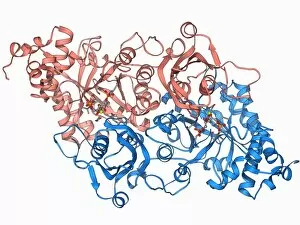
Background imageProteomics Collection: HIV-1 protease and inhibitor F006 / 9773

HIV-1 protease and inhibitor F006 / 9773
HIV-1 protease and inhibitor. Molecular model of the enzyme HIV-1 protease (pink and blue ribbons) bound to an inhibitor molecule (centre)

Background imageProteomics Collection: Ribonuclease A molecule F006 / 9768

Ribonuclease A molecule F006 / 9768
Ribonuclease A (RNAse A), molecular model. Ribonuclease (RNase) is a type of nuclease that catalyses the degradation of RNA (ribonucleic acid)

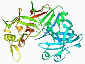
Background imageProteomics Collection: Pepsin stomach enzyme F006 / 9767

Pepsin stomach enzyme F006 / 9767
Pepsin stomach enzyme, molecular model. Pepsin is a protease enzyme that is secreted as part of gastric juice into the stomach in an inactive form known as pepsinogen

Background imageProteomics Collection: Flock house virus capsid F006 / 9755

Flock house virus capsid F006 / 9755
Flock house virus capsid, molecular model. The flock house virus is a member of the Nodaviridae family. It kills the New Zealand grass grub insect

Background imageProteomics Collection: Xylose isomerase complex F006 / 9765

Xylose isomerase complex F006 / 9765
Xylose isomerase complex. Molecular model of the enzyme D-xylose isomerase bound to the sugar alcohol sorbitol. D-xylose isomerase is involved in fructose and mannose metabolism

Background imageProteomics Collection: H-Ras p21 oncogene protein F006 / 9766

H-Ras p21 oncogene protein F006 / 9766
H-Ras p21 oncogene protein, molecular model. The Ras proteins are involved in transmitting signals within cells. Excessive signalling can lead to conditions such as cancer

Background imageProteomics Collection: Phosphofructokinase bacterial enzyme F006 / 9762

Phosphofructokinase bacterial enzyme F006 / 9762
Phosphofructokinase enzyme, molecular model. This enzyme, from the bacterium Bacillus stearothermophilus, is involved in regulating the process of releasing energy from glucose

Background imageProteomics Collection: tRNA molecule F006 / 9764

tRNA molecule F006 / 9764
Transfer RNA (tRNA), molecular model. tRNA (transfer ribonucleic acid) translates messenger RNA (mRNA) into a protein product

Background imageProteomics Collection: H-Ras p21 oncogene protein F006 / 9763

H-Ras p21 oncogene protein F006 / 9763
H-Ras p21 oncogene protein, molecular model. The Ras proteins are involved in transmitting signals within cells. Excessive signalling can lead to conditions such as cancer

Background imageProteomics Collection: Kinase inhibitor complex F006 / 9760

Kinase inhibitor complex F006 / 9760
Kinase inhibitor complex. Molecular model of a leucettine kinase inhibitor bound to a serine threonine kinase protein

Background imageProteomics Collection: Marburg viral protein 35 and RNA F006 / 9759

Marburg viral protein 35 and RNA F006 / 9759
Marburg viral protein 35 and RNA. Molecular model of the Marburg viral protein 35 (VP35) bound to a molecule of double stranded RNA (ribonucleic acid)

Background imageProteomics Collection: Insulin molecule F006 / 9761

Insulin molecule F006 / 9761
Insulin molecule. Molecular model of the hormone insulin from a pig. Insulin consists of two peptide chains, A and B, which are linked by disulphide bridges

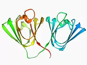
Background imageProteomics Collection: Eye lens protein molecule F006 / 9758

Eye lens protein molecule F006 / 9758
Eye lens protein. Molecular model of gammaB-crystallin, a protein found in the lens of the eye. The regular arrangement of the protein in the lens is thought to be responsible for its transparency

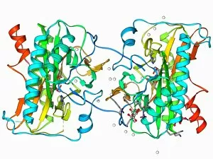
Background imageProteomics Collection: Methionine aminopeptidase molecule F006 / 9756

Methionine aminopeptidase molecule F006 / 9756
Methionine aminopeptidase, molecular model. This enzyme removes the amino acid methionine from proteins


All Professionally Made to Order for Quick Shipping

Proteomics, the study of proteins and their functions within an organism, is a fascinating field that unravels the intricate workings of life. From anaesthetics inhibiting ion channels to immunoglobulin G antibody molecules, proteomics delves into the molecular mechanisms that shape our existence. In the realm of brain research, scientists explore how proteins influence cognition and behavior. They investigate DNA nucleosomes' structure and function, unraveling their role in gene regulation. Antibodies take center stage as artwork showcases their diverse forms and crucial role in immune defense. Zinc fingers bound to a DNA strand highlight protein-DNA interactions critical for genetic processes. Meanwhile, manganese superoxide dismutase enzyme aids in protecting cells from oxidative stress. The SARS coronavirus protein becomes a subject of intense scrutiny as researchers strive to understand its pathogenicity. Cytochrome b5 molecule reveals insights into electron transfer reactions within cells while glutamine synthetase enzyme plays a vital role in nitrogen metabolism. Lastly, RNA-editing enzymes offer potential therapeutic targets for various diseases with their ability to modify genetic information at the RNA level. Through proteomics, we unlock nature's secrets one protein at a time - deciphering their structures, unraveling their functions, and ultimately enhancing our understanding of life itself.