Fraud Blocker Skip to main content

Proteomics Collection (page 6)

Proteomics, the study of proteins and their functions within an organism, is a fascinating field that unravels the intricate workings of life

Background imageProteomics Collection: Pepsinogen molecule F006 / 9710

Pepsinogen molecule F006 / 9710
Pepsinogen. Molecular model of pepsinogen, the inactive precursor to the digestive enzyme pepsin. Pepsion, which is released by the stomach digests proteins
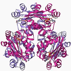
Background imageProteomics Collection: Glycosylation enzyme molecule F006 / 9708

Glycosylation enzyme molecule F006 / 9708
Glycosylation enzyme. Molecular model of the enzyme N-acetylglucosamine (GlcNAc) transferase. This intracellular enzyme adds N-acetylglucosamine molecules to target proteins

Background imageProteomics Collection: Retroviral intasome molecule F006 / 9705

Retroviral intasome molecule F006 / 9705
Retroviral intasome molecule. Molecular model of an intasome from a retrovirus complexed with host cell DNA (deoxyribonucleic acid)

Background imageProteomics Collection: Retroviral intasome molecule F006 / 9706

Retroviral intasome molecule F006 / 9706
Retroviral intasome molecule. Molecular model of an intasome from a retrovirus complexed with host cell DNA (deoxyribonucleic acid)

Background imageProteomics Collection: Metarhodopsin molecule F006 / 9709

Metarhodopsin molecule F006 / 9709
Metarhodopsin II molecule, molecular model. This is one of the intermediary breakdown products formed when light falls on the photoreceptor pigment rhodopsin

Background imageProteomics Collection: NpmA methyltransferase F006 / 9707

NpmA methyltransferase F006 / 9707
NpmA methyltransferase, molecular model. Methyltransferase enzymes act to add methyl groups to nucleic acids such as DNA (deoxyribonucleic acid), a process called DNA methylation

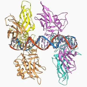
Background imageProteomics Collection: Notch transcription complex F006 / 9703

Notch transcription complex F006 / 9703
Notch transcription complex. Molecular model of the notch transcription complex bound to the human Hes1 promoter on a strand of DNA (deoxyribonucleic acid, red and blue)

Background imageProteomics Collection: Oxidoreductase enzyme complex F006 / 9700

Oxidoreductase enzyme complex F006 / 9700
Oxidoreductase enzyme complex. Molecular model of a complex of NADH-quinone oxidoreductase subunits. The whole is termed respiratory complex I

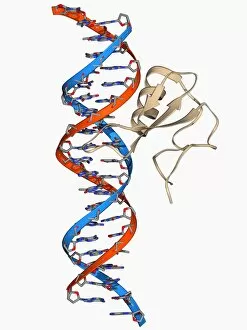
Background imageProteomics Collection: DNA repair enzyme, molecular model F006 / 9704

DNA repair enzyme, molecular model F006 / 9704
DNA repair enzyme. Molecular model of the DNA (deoxyribonucleic acid) repair enzyme dioxygenase bound to a molecule of DNA (red and blue)

Background imageProteomics Collection: Lassa virus nucleocapsid protein F006 / 9702

Lassa virus nucleocapsid protein F006 / 9702
Lassa virus nucleocapsid protein, molecular model. This protein has essential roles in RNA (ribonucleic acid) synthesis and immune suppression

Background imageProteomics Collection: Human rotavirus enterotoxin F006 / 9701

Human rotavirus enterotoxin F006 / 9701
Human rotavirus enterotoxin. Molecular model of the NSP4 (nonstructural protein 4) from the human rotavirus. This enterotoxin induces diarrhoea

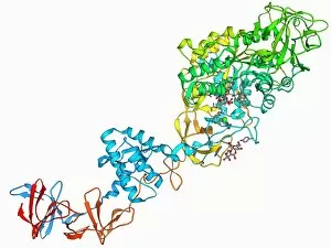
Background imageProteomics Collection: Isocitrate dehydrogenase kinase F006 / 9698

Isocitrate dehydrogenase kinase F006 / 9698
Isocitrate dehydrogenase kinase. Molecular model of isocitrate dehydrogenase kinase phosphatase (AceK) complexed with its substrate isocitrate dehydrogenase (ICDH)

Background imageProteomics Collection: Ebola viral protein 35 and RNA F006 / 9697

Ebola viral protein 35 and RNA F006 / 9697
Ebola viral protein 35 and RNA. Molecular model of the Ebola viral protein 35 (VP35) bound to a molecule of double stranded RNA (ribonucleic acid)

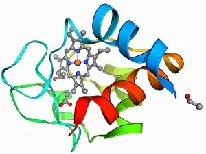
Background imageProteomics Collection: Cytochrome C, molecular model F006 / 9696

Cytochrome C, molecular model F006 / 9696
Cytochrome C, molecular model. Cytochrome molecules perform oxidation and reduction reactions for electron transport, a chain of reactions used to power cellular processes that require energy

Background imageProteomics Collection: Lactate dehydrogenase enzyme molecule F006 / 9699

Lactate dehydrogenase enzyme molecule F006 / 9699
Lactate dehydrogenase enzyme, molecular model. This enzyme converts pyruvate to lactate in the final steps of glycolysis, and lactate to pyruvate during the lactic acid cycle

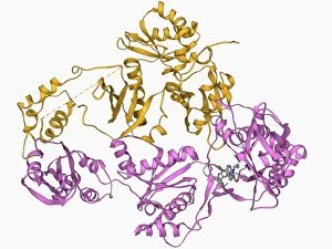
Background imageProteomics Collection: mRNA capping apparatus F006 / 9694

mRNA capping apparatus F006 / 9694
mRNA capping apparatus. Molecular model of the Cet-1-Ceg1 mRNA capping apparatus

Background imageProteomics Collection: Eye lens protein molecule F006 / 9695

Eye lens protein molecule F006 / 9695
Eye lens protein. Molecular model of alpha-crystallin, a protein found in the lens of the eye. The regular arrangement of the protein in the lens is thought to be responsible for its transparency

Background imageProteomics Collection: Transcription repressor protein and DNA F006 / 9692

Transcription repressor protein and DNA F006 / 9692
Transcription repressor protein and DNA, molecular model. The repressor protein (green) is binding to a strand of DNA (deoxyribonucleic acid, pink and purple)

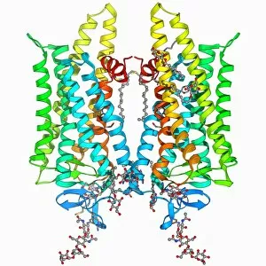
Background imageProteomics Collection: Zinc transporter Yiip molecule F006 / 9691

Zinc transporter Yiip molecule F006 / 9691
Zinc transporter Yiip, molecular model. This transmembrane protein facilitates the movement of zinc ions

Background imageProteomics Collection: Kinesin motor protein F006 / 9693

Kinesin motor protein F006 / 9693
Kinesin motor protein, molecular model. Kinesin motor proteins transport vesicles containing intracellular cargo around the cell along microtubules

Background imageProteomics Collection: West Nile virus and antibodies F006 / 9689

West Nile virus and antibodies F006 / 9689
West Nile virus and antibodies. Molecular model of antibodies attached to the capsid of a west Nile virus. West Nile Virus is an arbovirus (arthropod-borne virus)

Background imageProteomics Collection: Bluetongue virus capsid, molecular model F006 / 9687

Bluetongue virus capsid, molecular model F006 / 9687
Bluetongue virus capsid, molecular model. This virus, transmitted by midges, causes bluetongue disease in ruminants (commonly sheep, and less frequently cattle, goats and deer)

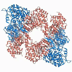
Background imageProteomics Collection: Chaperonin protein F006 / 9690

Chaperonin protein F006 / 9690
Chaperonin protein, molecular mode. Chaperonins are proteins that provide favourable conditions for the correct folding of other proteins

Background imageProteomics Collection: Hepatitis E virus capsid, molecular model F006 / 9688

Hepatitis E virus capsid, molecular model F006 / 9688
Hepatitis E virus capsid, molecular model. This virus causes the disease hepatitis E in humans. Transmitted by faecal-oral contact

Background imageProteomics Collection: Oestrogen receptor and tamoxifen drug F006 / 9674

Oestrogen receptor and tamoxifen drug F006 / 9674
Oestrogen receptor and tamoxifen drug, molecular model. Tamoxifen is a selective oestrogen receptor modulator (SERM) that blocks oestrogen production in breast tissue by inhibiting the oestrogen

Background imageProteomics Collection: Glucansucrase enzyme molecule F006 / 9685

Glucansucrase enzyme molecule F006 / 9685
Glucansucrase, molecular model. This enzyme catalyses the cleavage of sucrose into fructose and glucose

Background imageProteomics Collection: Cytochrome BC1 molecule F006 / 9681

Cytochrome BC1 molecule F006 / 9681
Cytochrome BC1, Molecular model. Cytochrome molecules perform oxidation and reduction reactions for electron transport, a chain of reactions used to power cellular processes that require energy

Background imageProteomics Collection: HIV reverse transcription enzyme F006 / 9684

HIV reverse transcription enzyme F006 / 9684
HIV reverse transcription enzyme. Molecular model of the reverse transcriptase enzyme found in HIV (the human immunodeficiency virus) bound to the inhibitor nevirapine

Background imageProteomics Collection: Human growth hormone molecule F006 / 9683

Human growth hormone molecule F006 / 9683
Human growth hormone. Molecular model of human growth hormone (hGH, orange) bound to the extracellular domain of the human growth hormone binding protein (hGHBP)

Background imageProteomics Collection: HIV DNA and transcription factor F006 / 9680

HIV DNA and transcription factor F006 / 9680
HIV DNA and transcription factor. Molecular model of DNA (deoxyribonucleic acid) from HIV-1 (human immunodeficiency virus type 1) complexed with the transcription factor kappa B

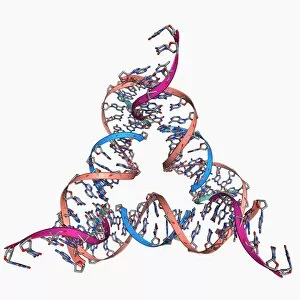
Background imageProteomics Collection: Self-assembled DNA triangle F006 / 9679

Self-assembled DNA triangle F006 / 9679
Self-assembled DNA triangle. Molecular model of DNA (deoxyribonucleic acid) strands forming what is called a tensegrity triangle

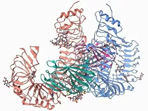
Background imageProteomics Collection: Human pathogen recognition molecule F006 / 9677

Human pathogen recognition molecule F006 / 9677
Human pathogen recognition molecule. Molecular model of a complex between human lymphocyte antigen 96 and toll-like receptor 4

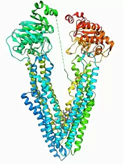
Background imageProteomics Collection: Antibodies and their antigen F006 / 9678

Antibodies and their antigen F006 / 9678
Antibodies and their antigen. Molecular model showing the molecular structure of two anti-P-glycoprotein antibodies (upper left and right) and the P-glycoprotein (v-shaped) to which they respond

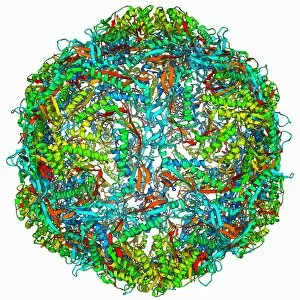
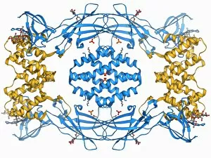
Background imageProteomics Collection: Bacterial nanocompartment F006 / 9672

Bacterial nanocompartment F006 / 9672
Bacterial nanocompartment. Molecular molecule of a nanocompartment from the bacterium Thermotoga maritima. This is a shell formed from encapsulin proteins that encloses enzymes that defend the cell

Background imageProteomics Collection: E coli virulence factor molecule F006 / 9675

E coli virulence factor molecule F006 / 9675
E. coli virulence factor molecule. Molecular model of the enzyme arylsulfate sulfotransferase (ASST) from an Escherichia coli bacterium

Background imageProteomics Collection: Fatty acid binding protein and inhibitor F006 / 9676

Fatty acid binding protein and inhibitor F006 / 9676
Fatty acid binding protein and inhibitor. Molecular model of adipocyte fatty-acid binding protein (A-FABP) bound to an inhibitor

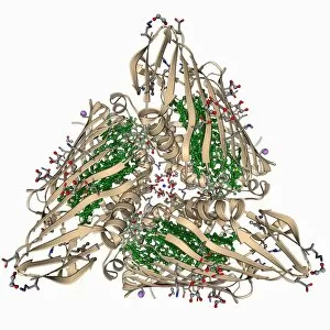
Background imageProteomics Collection: Fenna-Matthews-Olson complex molecule F006 / 9673

Fenna-Matthews-Olson complex molecule F006 / 9673
Fenna-Matthews-Olson complex. Molecular model of the Fenna-Matthews-Olson complex from the green sulphur bacterium Prosthecochloris aestuarii

Background imageProteomics Collection: Nucleoside diphosphate kinase molecule F006 / 9671

Nucleoside diphosphate kinase molecule F006 / 9671
Nucleoside diphosphate kinase. Molecular model of the enzyme nucleoside diphosphate kinase from the Acanthamoeba polyphaga mimivirus

Background imageProteomics Collection: Cytochrome C, molecular model F006 / 9670

Cytochrome C, molecular model F006 / 9670
Cytochrome C, molecular model. Cytochrome molecules perform oxidation and reduction reactions for electron transport, a chain of reactions used to power cellular processes that require energy

Background imageProteomics Collection: Concanavalin A F006 / 9668

Concanavalin A F006 / 9668
Concanavalin A, molecular model. This lectin protein is found in legumes. Lectins are highly specific sugar-binding proteins that play a role in biological recognition

Background imageProteomics Collection: Toll-like receptor 3 and RNA F006 / 9666

Toll-like receptor 3 and RNA F006 / 9666
Toll-like receptor 3 and RNA. Molecular model of the toll-like receptor 3 (TLR3) protein (pink and blue) bound to a strand of RNA (ribonucleic acid, green and yellow)

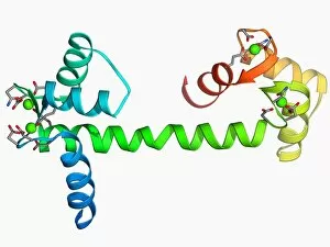
Background imageProteomics Collection: Calcium-binding protein molecule F006 / 9667

Calcium-binding protein molecule F006 / 9667
Calcium-binding protein. Molecule model of the calcium-binding protein calmodulin (CaM). This protein is found in all eukaryotic cells

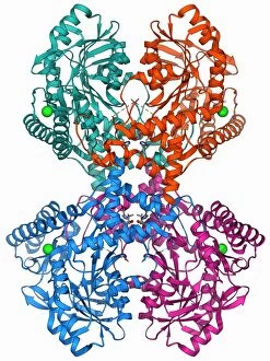
Background imageProteomics Collection: Isocitrate dehydrogenase molecule F006 / 9663

Isocitrate dehydrogenase molecule F006 / 9663
Isocitrate dehydrogenase, molecular model. This enzyme catalyses the third step in the citric acid (or Krebs) cycle, the process by which mitochondria convert glucose to energy

Background imageProteomics Collection: Opsin molecule F006 / 9665

Opsin molecule F006 / 9665
Opsin. Molecular model of a ligand-free opsin molecule. Opsins are found in photoreceptor cells (rods and cones) in the retina of the eye

Background imageProteomics Collection: Interferon antagonism by viral protein F006 / 9662

Interferon antagonism by viral protein F006 / 9662
Interferon (IFN) antagonism by viral protein. Molecular model of an orthopoxvirus IFN-gamma-binding protein bound to an IFN-gamma molecule

Background imageProteomics Collection: DNA and MECP2 complex, molecular model F006 / 9664

DNA and MECP2 complex, molecular model F006 / 9664
DNA and MECP2 complex. Molecular model of MECP2 (methyl CpG binding protein 2 (Rett syndrome)) bound to the BDNF (brain-derived neurotrophic factor)

Background imageProteomics Collection: Selenocysteine synthase enzyme molecule F006 / 9661

Selenocysteine synthase enzyme molecule F006 / 9661
Selenocysteine synthase enzyme molecule. Computer model showing the molecular structure of the enzyme selenocysteine synthase (SecS)

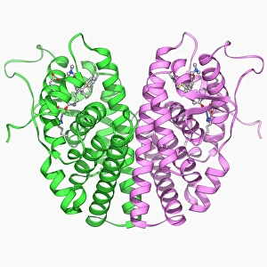
Background imageProteomics Collection: Androgen receptor and modulator F006 / 9660

Androgen receptor and modulator F006 / 9660
Androgen receptor and modulator. Molecular model of a selective androgen receptor modulator (SARM) bound to an androgen receptor


All Professionally Made to Order for Quick Shipping

Proteomics, the study of proteins and their functions within an organism, is a fascinating field that unravels the intricate workings of life. From anaesthetics inhibiting ion channels to immunoglobulin G antibody molecules, proteomics delves into the molecular mechanisms that shape our existence. In the realm of brain research, scientists explore how proteins influence cognition and behavior. They investigate DNA nucleosomes' structure and function, unraveling their role in gene regulation. Antibodies take center stage as artwork showcases their diverse forms and crucial role in immune defense. Zinc fingers bound to a DNA strand highlight protein-DNA interactions critical for genetic processes. Meanwhile, manganese superoxide dismutase enzyme aids in protecting cells from oxidative stress. The SARS coronavirus protein becomes a subject of intense scrutiny as researchers strive to understand its pathogenicity. Cytochrome b5 molecule reveals insights into electron transfer reactions within cells while glutamine synthetase enzyme plays a vital role in nitrogen metabolism. Lastly, RNA-editing enzymes offer potential therapeutic targets for various diseases with their ability to modify genetic information at the RNA level. Through proteomics, we unlock nature's secrets one protein at a time - deciphering their structures, unraveling their functions, and ultimately enhancing our understanding of life itself.