Fraud Blocker Skip to main content

Compounds Collection (page 9)

"Exploring the Fascinating World of Compounds: From Copper and Magnesium Sulphate to Graphene" Delving into the intricate beauty of compounds

Background imageCompounds Collection: Wolcott Gibbs, US chemist

Wolcott Gibbs, US chemist
Wolcott Gibbs (1822-1908), US chemist. Gibbs graduated from Columbia University, USA in 1841 and qualified as a medical doctor in 1845

Background imageCompounds Collection: Carbon nanotube and buckyball, artwork

Carbon nanotube and buckyball, artwork
Carbon nanotube and buckyball. Computer artwork showing the hexagonal carbon structures of a buckyball (yellow) inside a carbon nanotube, or buckytube (grey)

Background imageCompounds Collection: Levitra drug molecule

Levitra drug molecule
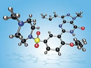
Levitra. Molecular model of the drug vardenafil, marketed under the brand Levitra. Atoms are represented as spheres and are colour-coded: carbon (black), hydrogen (silver), nitrogen (blue)

Background imageCompounds Collection: Cialis drug molecule

Cialis drug molecule
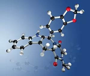
Cialis. Molecular model of the drug tadalafil, marketed under the brand Cialis. Atoms are represented as spheres and are colour-coded: carbon (black), hydrogen (grey), nitrogen (blue), oxygen (red)

Background imageCompounds Collection: Fluoxetine drug molecule

Fluoxetine drug molecule
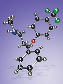
Fluoxetine. Molecular model of the drug fluoxetine, which is marketed under the brand Prozac. Atoms are represented as spheres and are colour-coded: carbon (black), hydrogen (silver)

Background imageCompounds Collection: Viagra drug molecule

Viagra drug molecule
Viagra. Molecular model of the drug sildenafil citrate, marketed under the brands Viagra and Revatio. Atoms are represented as spheres and are colour-coded: carbon (black), hydrogen (silver)

Background imageCompounds Collection: Synthetic fibre, light micrograph

Synthetic fibre, light micrograph
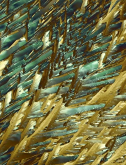
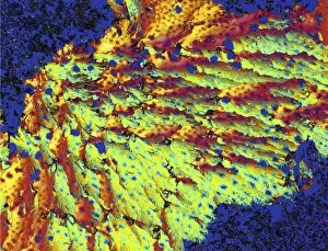
Light micrograph of a synthetic fibre, taken with polarised light. Magnification: x100

Background imageCompounds Collection: Glycine crystals, light micrograph

Glycine crystals, light micrograph
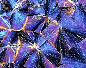
Glycine crystals. Polarised light micrograph of crystals of glycine, the simplest of the amino acids. Glycine consists of a central carbon atom, to which are attached two hydrogen atoms

Background imageCompounds Collection: Adrenaline, polarized light micrograph

Adrenaline, polarized light micrograph
Adrenaline. Polarized light micrograph of crystals of adrenaline (epinephrine). This is a hormone produced in the adrenal glands above the kidneys

Background imageCompounds Collection: Serotonin crystals, light micrograph

Serotonin crystals, light micrograph
Serotonin. Polarised light micrograph of serotonin, a neurotransmitter. Serotonin is derived from tryptophan, and is found in the vertebrate brain

Background imageCompounds Collection: Ice, molecular structure

Ice, molecular structure
Ice. Molecular model showing the hexagonal structure of ice. Atoms are represented as spheres and are colour-coded: hydrogen (blue) and oxygen (red). The lattice structure of ice gives it its rigidity

Background imageCompounds Collection: Myoglobin molecule

Myoglobin molecule. Computer model showing the structure of a Myoglobin molecule. Myoglobin is a protein found in muscle tissue

Background imageCompounds Collection: Cyclodextrin molecule, artwork

Cyclodextrin molecule, artwork
Cyclodextrin. Molecular model of beta-cyclodextrin, which comprises seven sugar molecules bound in a ring. Cyclodextrins are used in the food

Background imageCompounds Collection: Development of an atheroma, artwork

Development of an atheroma, artwork
Cyclodextrins. Molecular models of alpha- (left), beta- (centre) and gamma- (right) cyclodextrin molecules. These comprise of six (alpha), seven (beta)

Background imageCompounds Collection: Hippuric acid crystals, light micrograph

Hippuric acid crystals, light micrograph
Polarised light micrograph of Hippuric Acid crystals. Hippuric acid is one of the purine family of amino-acids, and is also known as Benzoylaminoacetic Acid or N-benzoylglycine

Background imageCompounds Collection: Insulin molecule, close-up view

Insulin molecule, close-up view
Insulin. Computer graphic showing a part of the molecule of human insulin. Insulin is a hormone which is produced in the pancreas by cells of the Islets of Langerhans

Background imageCompounds Collection: Tartaric acid crystals, light micrograph

Tartaric acid crystals, light micrograph
Polarised light micrograph of crystals of tartaric acid. Tartaric acid is used in baking powders & as a food additive. It can be obtained from tartar (potassium hydrogen tartrate)

Background imageCompounds Collection: Insulin crystals, light micrograph

Insulin crystals, light micrograph
Insulin. Polarised light micrograph (PLM) of crystals of the hormone insulin. The crystals appear hexagonal in shape. The insulin molecule is made up of two chains of amino acids (A & B chains)

Background imageCompounds Collection: Progesterone hormone, light micrograph

Progesterone hormone, light micrograph
Progesterone hormone. Polarised light micrograph of crystals of female sex hormone progesterone. Progesterone is the most potent of the naturally- occurring progestogens, a class of steroid hormones

Background imageCompounds Collection: Nikolai Vorozhtsov, Russian chemist

Nikolai Vorozhtsov, Russian chemist
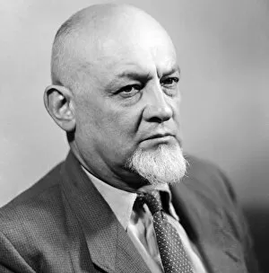
Soviet academician Nikolai Vorozhtsov

Background imageCompounds Collection: Human ApolipoproteinA-1

Human ApolipoproteinA-1
Molecular model of high-density lipoprotein (HDL) known as an apolipoprotein. HDLs are proteins that bind to lipid (fat) molecules and transport them around the body

Background imageCompounds Collection: Search for a new drug, conceptual image

Search for a new drug, conceptual image

Background imageCompounds Collection: Carbon nanotube, artwork

Carbon nanotube, artwork
Carbon nanotube. Computer artwork showing the hexagonal carbon structure of a nanotube, or buckytube

Background imageCompounds Collection: Graphene, molecular structure

Graphene, molecular structure

Background imageCompounds Collection: Silver Nitrate, light micrograph

Silver Nitrate, light micrograph
Composite crystals of Silver Nitrate, polarised light micrograph

Background imageCompounds Collection: Lysyl oxidase enzyme molecule

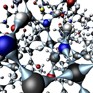
Lysyl oxidase enzyme molecule. Computer artwork showing the secondary structure of the enzyme lysyl oxidase (LOX). LOX is a homodimeric (composed of two identical subunits)

Background imageCompounds Collection: Hantavirus inhibitor molecule

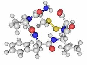
Hantavirus inhibitor molecule. Molecular model of a pentapeptide protein that blocks the entry of hantavirus particles to human cells

Background imageCompounds Collection: Anticoagulant molecule

Anticoagulant molecule. Molecular model of one subunit of the anticoagulant heparin. Atoms are represented as spheres and are colour-coded: carbon (green), hydrogen (white), oxygen (red)

Background imageCompounds Collection: DEET insect repellent molecule

DEET insect repellent molecule
DEET. Molecular model of the insect repellent DEET (diethy-meta-toluamide). DEET is the most common active ingredient used in insect repellents. It is primarily used to repel mosquitoes and ticks


All Professionally Made to Order for Quick Shipping

"Exploring the Fascinating World of Compounds: From Copper and Magnesium Sulphate to Graphene" Delving into the intricate beauty of compounds, we witness the mesmerizing sight of copper and magnesium sulphate crystals under a light microscope (LM). A closer look at caffeine crystals through a light micrograph reveals their captivating structure, resembling tiny jewels that fuel our mornings. Oxytocin hormone crystals, captured using polarized light microscopy (PLM C016 / 7196), unveil the remarkable complexity behind this molecule responsible for human bonding. Through an artistic representation, we unravel the secondary structure of proteins – nature's building blocks that orchestrate countless biological processes within us. The perovskite crystal structure captivates scientists with its potential applications in renewable energy technologies, promising a brighter future for sustainable power generation. Another glimpse into oxytocin's world showcases its crystalline form under a light microscope, reminding us of its vital role in nurturing social connections and maternal instincts. Zooming in on caffeine's molecular composition unveils its drug-like qualities that stimulate our nervous system and keep us awake during long nights or early mornings. Peering into the microscopic realm reveals bacterial ribosomes - miniature protein factories essential for life itself - showcasing nature's incredible machinery at work. Cortisol crystals come to life as they are illuminated by a beam of light under a microscope, offering insight into this stress hormone's unique properties within our bodies. Exploring vitamin B12 through its molecular model highlights how this crucial nutrient supports various bodily functions while displaying an elegant arrangement of atoms and bonds. Once again, copper sulphate crystals enchant us with their vibrant colors when observed using a light microscope (LM), reminding us of their diverse industrial uses and chemical significance. Stepping into the realm of materials science brings forth graphene.