Fraud Blocker Skip to main content

Compounds Collection (page 4)

"Exploring the Fascinating World of Compounds: From Copper and Magnesium Sulphate to Graphene" Delving into the intricate beauty of compounds

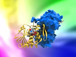
Background imageCompounds Collection: Ricin molecule, artwork C017 / 3656

Ricin molecule, artwork C017 / 3656
Ricin molecule. Computer artwork showing the structure of a molecule of the toxic protein ricin. Ricin comprises two entwined amino acid chains; A (yellow) and B (blue)

Background imageCompounds Collection: Water molecules, artwork C017 / 7384

Water molecules, artwork C017 / 7384
Water molecules. Computer artwork showing the molecular (top) and atomic (bottom) structure of water (H2O). Atoms are colour-coded: hydrogen (blue) and oxygen (white)

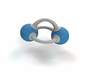
Background imageCompounds Collection: Methane molecule, artwork C017 / 3614

Methane molecule, artwork C017 / 3614
Methane molecule. Computer artwork showing the structure of a molecule of nitrogen (N2). Atoms are colour coded: nitrogen (blue), with the bonds between them as bars (grey)

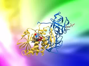
Background imageCompounds Collection: Ricin molecule, artwork C017 / 3655

Ricin molecule, artwork C017 / 3655
Ricin molecule. Computer artwork showing the structure of a molecule of the toxic protein ricin. Ricin comprises two entwined amino acid chains; A (yellow) and B (blue)

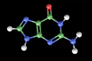
Background imageCompounds Collection: Guanine molecule, artwork C017 / 7239

Guanine molecule, artwork C017 / 7239
Guanine molecule. Computer artwork showing the structure of a molecule of the nucleobase guanine. Atoms are shown as colour-coded spheres: carbon (green), hydrogen (white)

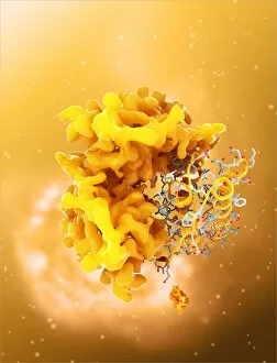
Background imageCompounds Collection: Ricin molecule, artwork C017 / 3648

Ricin molecule, artwork C017 / 3648
Ricin molecule Computer artwork showing the structure of a molecule of the toxic protein ricin (blue and yellow) with an active ribosome in the background

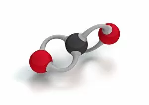
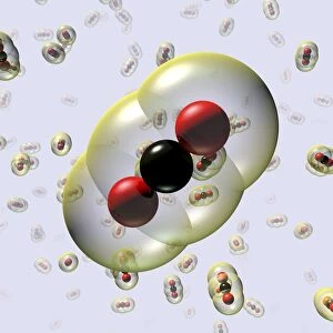
Background imageCompounds Collection: Carbon dioxide molecule C017 / 3600

Carbon dioxide molecule C017 / 3600
Carbon dioxide molecule. Computer artwork showing the structure of a molecule of carbon dioxide (CO2). Atoms are colour coded: oxygen (red) and carbon (black)

Background imageCompounds Collection: Thymine-adenine interaction, artwork C017 / 7368

Thymine-adenine interaction, artwork C017 / 7368
Thymine-adenine interaction. Computer artwork showing the structure of bound thymine and adenine molecules. Atoms are shown as colour-coded spheres: carbon (green), hydrogen (white)

Background imageCompounds Collection: Resveratrol molecule C014 / 2110

Resveratrol molecule C014 / 2110
Resveratrol, molecular model. Resveratrol is a phytoalexin, a defence chemical produced by plants, found in grapes, peanuts, blueberries and some pines

Background imageCompounds Collection: Cytosine molecule, artwork C017 / 7213

Cytosine molecule, artwork C017 / 7213
Cytosine molecule. Computer artwork showing the structure of a molecule of the nucleobase cytosine (2-oxy-4-aminopyrimidine)

Background imageCompounds Collection: Rotaxane, molecular crystal structure C017 / 7010

Rotaxane, molecular crystal structure C017 / 7010
Molecular crystal structure of a rotaxane. A rotaxane is a chemical compound composed of a linear molecular chain passing through a chainlike molecular ring

Background imageCompounds Collection: Guanine molecule, artwork C017 / 7238

Guanine molecule, artwork C017 / 7238
Guanine molecule. Computer artwork showing the structure of a molecule of the nucleobase guanine. Atoms are shown as colour-coded spheres: carbon (green), hydrogen (white)

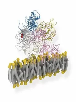
Background imageCompounds Collection: Coagulation factor complex molecule C014 / 0409

Coagulation factor complex molecule C014 / 0409
Coagulation factor complex molecule. Molecular model showing the interaction between coagulation factor VIII (FVIII, pink, blue and yellow), factor IXa (FIXa)

Background imageCompounds Collection: Insulin molecule C014 / 2122

Insulin molecule C014 / 2122
Insulin, molecular module. Insulin is a hormone produced by the pancreas. It consists of two peptide chains, A (blue) and B (yellow), which are linked by disulphide bridges

Background imageCompounds Collection: Resveratrol molecule C014 / 2108

Resveratrol molecule C014 / 2108
Resveratrol, molecular model. Resveratrol is a phytoalexin, a defence chemical produced by plants, found in grapes, peanuts, blueberries and some pines

Background imageCompounds Collection: Insulin molecule C014 / 2290

Insulin molecule C014 / 2290
Insulin molecule. Molecular module of insulin showing its primary structure over a background of the molecules electron density map

Background imageCompounds Collection: Resveratrol molecule C014 / 2109

Resveratrol molecule C014 / 2109
Resveratrol, molecular model. Resveratrol is a phytoalexin, a defence chemical produced by plants, found in grapes, peanuts, blueberries and some pines

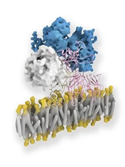
Background imageCompounds Collection: Coagulation factor complex molecule C014 / 0410

Coagulation factor complex molecule C014 / 0410
Coagulation factor complex molecule. Molecular model showing the interaction between coagulation factor VIII (FVIII, pink, blue and yellow), factor IXa (FIXa)

Background imageCompounds Collection: Stilbene synthase molecule C014 / 2292

Stilbene synthase molecule C014 / 2292
Stilbene synthase, molecular model. This enzyme is produced by some plants (including grapes, peanuts and blueberries) in response to stress from either ultraviolet light or certain fungi

Background imageCompounds Collection: Insulin molecule C014 / 2120

Insulin molecule C014 / 2120
Insulin molecule. Molecular module of insulin showing its primary structure. Insulin is a hormone produced by the pancreas

Background imageCompounds Collection: Stilbene synthase molecule C014 / 2291

Stilbene synthase molecule C014 / 2291
Stilbene synthase, molecular model. This enzyme is produced by some plants (including grapes, peanuts and blueberries) in response to stress from either ultraviolet light or certain fungi

Background imageCompounds Collection: UVR8 protein molecule C014 / 4913

UVR8 protein molecule C014 / 4913
UVR8 protein molecule. Computer model showing photoreception of UV-B (ultraviolet-B) light rays (white beam, left) by a UVR8 protein, whose secondary structure (purple ribbons) is shown

Background imageCompounds Collection: Leptin molecule C014 / 4904

Leptin molecule C014 / 4904
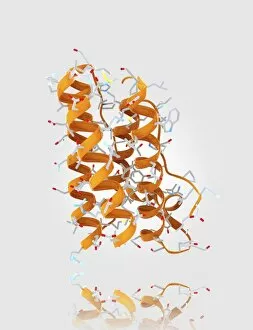
Leptin molecule. Computer model showing the structure of a molecule of the hormone leptin. Leptin is produced by adipose (fat) tissue

Background imageCompounds Collection: Peptide YY obesity hormone molecule C014 / 4911

Peptide YY obesity hormone molecule C014 / 4911
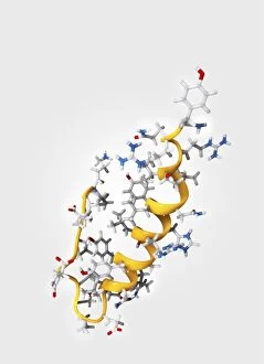
Peptide YY obesity hormone molecule. Computer model showing the crystal structure of a molecule of the hormone peptide YY (PYY, or PYY3-36)

Background imageCompounds Collection: Peptide YY obesity hormone molecule C014 / 4910

Peptide YY obesity hormone molecule C014 / 4910
Peptide YY obesity hormone molecule. Computer model showing the crystal structure of a molecule of the hormone peptide YY (PYY, or PYY3-36)

Background imageCompounds Collection: Leptin molecule C014 / 4907

Leptin molecule C014 / 4907
Leptin molecule. Computer model showing the crystal structure of a molecule of the hormone leptin. Leptin is produced by adipose (fat) tissue

Background imageCompounds Collection: Methyl sulphonal crystals, micrograph

Methyl sulphonal crystals, micrograph
Methyl sulphonal crystals, polarised light micrograph. Magnification: x114 when printed 10 centimetres wide

Background imageCompounds Collection: Activated ghrelin hormone molecule C014 / 4902

Activated ghrelin hormone molecule C014 / 4902
Activated ghrelin hormone molecule. Computer model showing the crystal structure of the human hormone ghrelin. The crystal structure consists of both the secondary structure

Background imageCompounds Collection: Activated ghrelin hormone molecule C014 / 4903

Activated ghrelin hormone molecule C014 / 4903
Activated ghrelin hormone molecule. Computer model showing the structure of the human hormone ghrelin. Atoms are colour-coded spheres (carbon: grey, oxygen: red, nitrogen: blue)

Background imageCompounds Collection: Leptin molecule and fat cells C014 / 4906

Leptin molecule and fat cells C014 / 4906
Leptin molecule and fat cells. Computer artwork showing a molecule of the hormone leptin with adipose (fat) cells (round), from which it is produced

Background imageCompounds Collection: Obestatin molecule C014 / 4908

Obestatin molecule C014 / 4908
Obestatin molecule. Computer artwork showing the structure of a molecule of obestatin. Obestatin is thought to supress hunger and reduce food intake, thereby reducing weight gain

Background imageCompounds Collection: Obestatin molecule C014 / 4909

Obestatin molecule C014 / 4909
Obestatin molecule. Computer artwork showing the structure of a molecule of obestatin. Obestatin is thought to supress hunger and reduce food intake, thereby reducing weight gain

Background imageCompounds Collection: UVR8 protein molecule C014 / 4912

UVR8 protein molecule C014 / 4912
UVR8 protein molecule. Computer model showing photoreception of UV-B (ultraviolet-B) light rays (top) by a UVR8 protein. The secondary structure (purple ribbons) of UVR8 is shown at bottom

Background imageCompounds Collection: Activated ghrelin hormone molecule C014 / 4901

Activated ghrelin hormone molecule C014 / 4901
Activated ghrelin hormone molecule. Computer model showing the crystal structure of the human hormone ghrelin. The crystal structure consists of both the secondary structure

Background imageCompounds Collection: Paracetamol molecule

Paracetamol molecule
Serotonin molecule. Computer model showing the structure of a molecule of the neurotransmitter (nerve signalling chemical) serotonin (5-hydroxytryptamine)

Background imageCompounds Collection: Water molecules C016 / 8541

Water molecules C016 / 8541
Water molecules. Molecular model of molecules of water. Water is one of the most abundant chemicals on Earth, covering almost 75 per cent of its surface

Background imageCompounds Collection: Water molecules C016 / 8540

Water molecules C016 / 8540
Water molecules. Molecular model of molecules of water. Water is one of the most abundant chemicals on Earth, covering almost 75 per cent of its surface

Background imageCompounds Collection: Water molecules C016 / 8538

Water molecules C016 / 8538
Water molecules. Molecular model of molecules of water. Water is one of the most abundant chemicals on Earth, covering almost 75 per cent of its surface

Background imageCompounds Collection: Water molecules C016 / 8537

Water molecules C016 / 8537
Water molecules. Molecular model of molecules of water. Water is one of the most abundant chemicals on Earth, covering almost 75 per cent of its surface

Background imageCompounds Collection: Water molecules C016 / 8536

Water molecules C016 / 8536
Water molecules. Molecular model of molecules of water. Water is one of the most abundant chemicals on Earth, covering almost 75 per cent of its surface

Background imageCompounds Collection: Water molecule C016 / 8535

Water molecule C016 / 8535
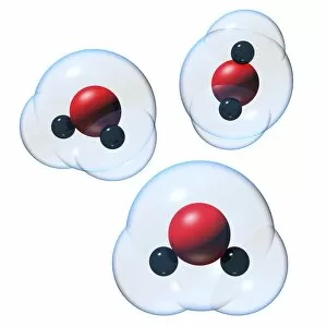
Water molecule. Molecular model of a molecule of water. Water is one of the most abundant chemicals on Earth, covering almost 75 per cent of its surface

Background imageCompounds Collection: Water molecule C016 / 8534

Water molecule C016 / 8534
Water molecule. Molecular model of a molecule of water. Water is one of the most abundant chemicals on Earth, covering almost 75 per cent of its surface

Background imageCompounds Collection: Graphene, molecular structure C016 / 8518

Graphene, molecular structure C016 / 8518
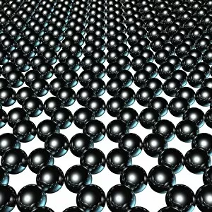
Graphene. Computer model of the molecular structure of graphene, a single layer of graphite. It is composed of hexagonally arranged carbon atoms (black) linked by strong covalent bonds (pink)
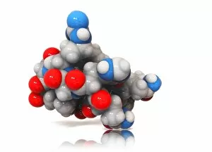
Background imageCompounds Collection: Graphene, molecular structure C016 / 8517

Graphene, molecular structure C016 / 8517
Graphene. Computer model of the molecular structure of graphene, a single layer of graphite. It is composed of hexagonally arranged carbon atoms (black) linked by strong covalent bonds (grey)
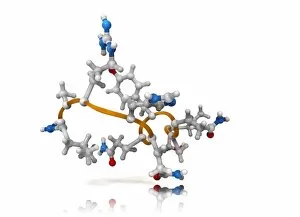
Background imageCompounds Collection: Graphene, molecular structure C016 / 8515

Graphene, molecular structure C016 / 8515
Graphene. Computer model of the molecular structure of graphene, a single layer of graphite. It is composed of hexagonally arranged carbon atoms (spheres) linked by strong covalent bonds

Background imageCompounds Collection: Graphene, molecular structure C016 / 8513

Graphene, molecular structure C016 / 8513
Graphene. Computer model of the molecular structure of graphene, a single layer of graphite. It is composed of hexagonally arranged carbon atoms (spheres) linked by strong covalent bonds (rods)

Background imageCompounds Collection: Graphene, molecular structure C016 / 8509

Graphene, molecular structure C016 / 8509
Graphene. Computer model of the molecular structure of graphene, a single layer of graphite. It is composed of hexagonally arranged carbon atoms (spheres) linked by strong covalent bonds

Background imageCompounds Collection: Carbon dioxide molecules C016 / 8495

Carbon dioxide molecules C016 / 8495
Carbon dioxide molecules. Computer artwork showing the structure of a molecule of carbon dioxide. Carbon dioxide is a colourless gas that occurs naturally in the atmosphere


All Professionally Made to Order for Quick Shipping

"Exploring the Fascinating World of Compounds: From Copper and Magnesium Sulphate to Graphene" Delving into the intricate beauty of compounds, we witness the mesmerizing sight of copper and magnesium sulphate crystals under a light microscope (LM). A closer look at caffeine crystals through a light micrograph reveals their captivating structure, resembling tiny jewels that fuel our mornings. Oxytocin hormone crystals, captured using polarized light microscopy (PLM C016 / 7196), unveil the remarkable complexity behind this molecule responsible for human bonding. Through an artistic representation, we unravel the secondary structure of proteins – nature's building blocks that orchestrate countless biological processes within us. The perovskite crystal structure captivates scientists with its potential applications in renewable energy technologies, promising a brighter future for sustainable power generation. Another glimpse into oxytocin's world showcases its crystalline form under a light microscope, reminding us of its vital role in nurturing social connections and maternal instincts. Zooming in on caffeine's molecular composition unveils its drug-like qualities that stimulate our nervous system and keep us awake during long nights or early mornings. Peering into the microscopic realm reveals bacterial ribosomes - miniature protein factories essential for life itself - showcasing nature's incredible machinery at work. Cortisol crystals come to life as they are illuminated by a beam of light under a microscope, offering insight into this stress hormone's unique properties within our bodies. Exploring vitamin B12 through its molecular model highlights how this crucial nutrient supports various bodily functions while displaying an elegant arrangement of atoms and bonds. Once again, copper sulphate crystals enchant us with their vibrant colors when observed using a light microscope (LM), reminding us of their diverse industrial uses and chemical significance. Stepping into the realm of materials science brings forth graphene.