Fraud Blocker Skip to main content

Compounds Collection (page 3)

"Exploring the Fascinating World of Compounds: From Copper and Magnesium Sulphate to Graphene" Delving into the intricate beauty of compounds

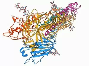
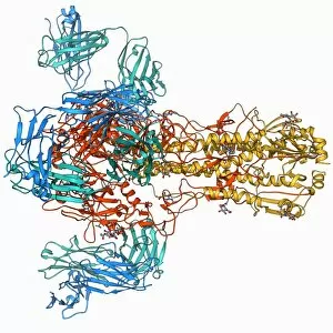
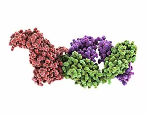
Background imageCompounds Collection: Haemagglutinin viral surface protein F007 / 9931

Haemagglutinin viral surface protein F007 / 9931
Haemagglutinin viral surface protein. Molecular model of haemagglutinin, a surface protein from the influenza virus, complexed with a neutralising antibody

Background imageCompounds Collection: Iron containing protein, molecular model

Iron containing protein, molecular model
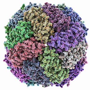
Iron containing protein. Molecular model showing the structure of a bacterial homolog of the animal iron containing protein ferritin

Background imageCompounds Collection: Proliferating cell nuclear antigen

Proliferating cell nuclear antigen molecule. Molecular model of human proliferating cell nuclear antigen (PCNA, blue, green and red), complexed with its loader protein (purple, orange)

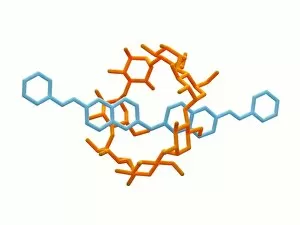
Background imageCompounds Collection: Rotaxane, molecular crystal structure C017 / 7011

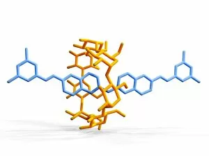
Rotaxane, molecular crystal structure C017 / 7011
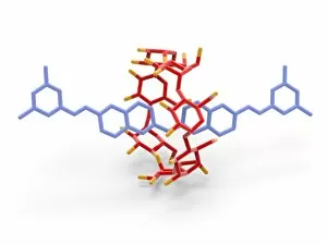
Molecular crystal structure of a rotaxane. A rotaxane is a chemical compound composed of a linear molecular chain passing through a chainlike molecular ring
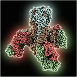
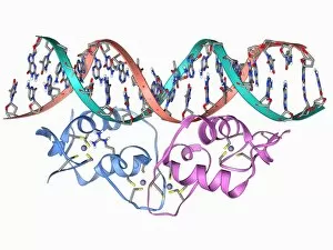
Background imageCompounds Collection: Tumour suppressor protein and DNA C017 / 3645

Tumour suppressor protein and DNA C017 / 3645
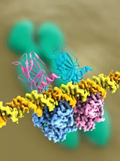
Tumour suppressor protein and DNA. Computer artwork showing a molecule of the tumour suppressor protein p53 (blue and pink) bound to a molecule of DNA (deoxyribonucleic acid, yellow and orange)

Background imageCompounds Collection: Organic chemistry building blocks C017 / 3599

Organic chemistry building blocks C017 / 3599
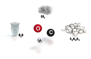
Organic chemistry building blocks, conceptual image. Computer artwork showing how carbon (C, black) oxygen (O, red) and hydrogen (H)

Background imageCompounds Collection: Ricin A-chain, artwork C017 / 3654

Ricin A-chain, artwork C017 / 3654
Ricin A-chain. Computer artwork showing the enzymatically active A-chain from a molecule of the toxic protein ricin. Ricin comprises two entwined amino acid chains; A (seen here) and B (not shown)
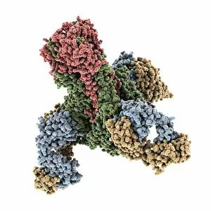
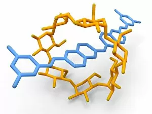
Background imageCompounds Collection: Rotaxane, molecular crystal structure C017 / 7014

Rotaxane, molecular crystal structure C017 / 7014
Molecular crystal structure of a rotaxane. A rotaxane is a chemical compound composed of a linear molecular chain passing through a chainlike molecular ring

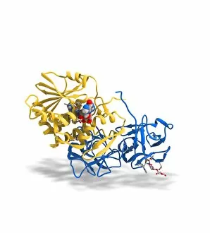
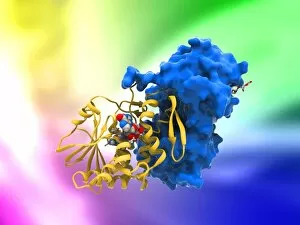
Background imageCompounds Collection: Ricin molecule, artwork C017 / 3649

Ricin molecule, artwork C017 / 3649
Ricin molecule. Computer artwork showing the structure of a molecule of the toxic protein ricin. Ricin comprises two entwined amino acid chains; A (yellow) and B (blue)

Background imageCompounds Collection: Rotaxane, molecular crystal structure C017 / 7013

Rotaxane, molecular crystal structure C017 / 7013
Molecular crystal structure of a rotaxane. A rotaxane is a chemical compound composed of a linear molecular chain passing through a chainlike molecular ring

Background imageCompounds Collection: Insulin molecule C014 / 2121

Insulin molecule C014 / 2121
Insulin molecule. Molecular module of insulin showing its secondary structure. Insulin is a hormone produced by the pancreas

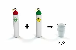
Background imageCompounds Collection: Reaction of hydrogen and oxygen to water C017 / 3607

Reaction of hydrogen and oxygen to water C017 / 3607
Reaction of hydrogen and oxygen to water. Computer artwork showing how hydrogen (H2, left) combines with oxygen (O2, centre) to form two water (H2O, right)

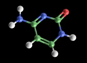
Background imageCompounds Collection: Cytosine molecule, artwork C017 / 7214

Cytosine molecule, artwork C017 / 7214
Cytosine molecule. Computer artwork showing the structure of a molecule of the nucleobase cytosine (2-oxy-4-aminopyrimidine)

Background imageCompounds Collection: H5N1 Haemagglutinin protein subunit F006 / 9590

H5N1 Haemagglutinin protein subunit F006 / 9590
H5N1 haemagglutinin protein subunit. Molecular model of the haemagglutinin HA(5) subunit. Haemagglutinin is a surface protein from the influenza A virus

Background imageCompounds Collection: Rotaxane, molecular crystal structure C017 / 7015

Rotaxane, molecular crystal structure C017 / 7015
Molecular crystal structure of a rotaxane. A rotaxane is a chemical compound composed of a linear molecular chain passing through a chainlike molecular ring

Background imageCompounds Collection: HIV reverse transcription enzyme F006 / 9494

HIV reverse transcription enzyme F006 / 9494
HIV reverse transcription enzyme. Molecular model of the reverse transcriptase enzyme (blue and green) found in HIV (the human immunodeficiency virus)

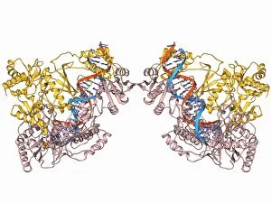
Background imageCompounds Collection: Transcription factor and DNA molecule F006 / 9484

Transcription factor and DNA molecule F006 / 9484
Transcription factor and DNA molecule. Molecular model of glucocorticoid receptor (GR) transcription factor protein (pink and blue) complexed with a molecule of DNA (deoxyribonucleic acid)

Background imageCompounds Collection: Haemagglutinin protein subunit F006 / 9479

Haemagglutinin protein subunit F006 / 9479
Haemagglutinin protein subunit. Molecular model of the ectodomain of the haemagglutinin HA(2) subunit. Haemagglutinin is a surface protein from the influenza A virus

Background imageCompounds Collection: Haemagglutinin viral surface protein F006 / 9470

Haemagglutinin viral surface protein F006 / 9470
Haemagglutinin viral surface protein. Molecular model of haemagglutinin, a surface protein from the influenza virus, complexed with a neutralising antibody

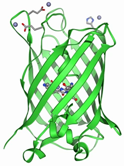
Background imageCompounds Collection: Green fluorescent protein molecule F006 / 9402

Green fluorescent protein molecule F006 / 9402
Green fluorescent protein (GFP), molecular model. The molecule has a cylindrical structure formed from beta sheets (ribbons). GFP is found in the Pacific jellyfish Aequorea victoria

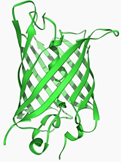
Background imageCompounds Collection: Green fluorescent protein molecule F006 / 9343

Green fluorescent protein molecule F006 / 9343
Green fluorescent protein (GFP), molecular model. The molecule has a cylindrical structure formed from beta sheets (ribbons). GFP is found in the Pacific jellyfish Aequorea victoria

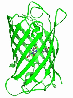
Background imageCompounds Collection: Green fluorescent protein molecule F006 / 9313

Green fluorescent protein molecule F006 / 9313
Green fluorescent protein (GFP), molecular model. The molecule has a cylindrical structure formed from beta sheets (ribbons). GFP is found in the Pacific jellyfish Aequorea victoria

Background imageCompounds Collection: Rotaxane, molecular crystal structure C017 / 7016

Rotaxane, molecular crystal structure C017 / 7016
Molecular crystal structure of a rotaxane. A rotaxane is a chemical compound composed of a linear molecular chain passing through a chainlike molecular ring

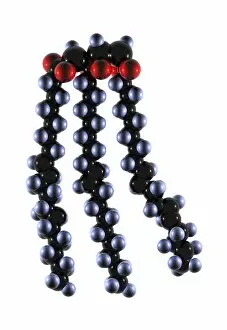
Background imageCompounds Collection: Polyunsaturated fat molecule

Polyunsaturated fat molecule. Computer model showing the structure of three unsaturated fat molecules bound together to form a polyunsaturated fat molecule

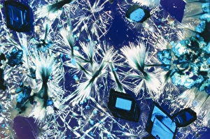
Background imageCompounds Collection: Copper sulphate crystals light micrograph

Copper sulphate crystals light micrograph
Copper sulphate crystals, polarised light micrograph. Magnification: x2.5, when printed 10 centimetres wide

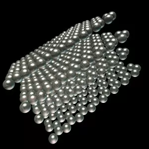
Background imageCompounds Collection: Graphite, molecular model C016 / 8894

Graphite, molecular model C016 / 8894
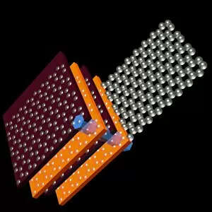
Graphite, molecular model. Graphite is used in pencil leads and as a lubricant. It is composed of parallel layers of hexagonally arranged carbon atoms (spheres)

Background imageCompounds Collection: Graphite, molecular model C016 / 8895

Graphite, molecular model C016 / 8895
Graphite, molecular model. Graphite is used in pencil leads and as a lubricant. It is composed of parallel layers of hexagonally arranged carbon atoms (spheres)

Background imageCompounds Collection: Graphite, molecular model C016 / 8896

Graphite, molecular model C016 / 8896
Graphite, molecular model. Graphite is used in pencil leads and as a lubricant. It is composed of parallel layers of hexagonally arranged carbon atoms (spheres)

Background imageCompounds Collection: Bionic nanotechnology, conceptual artwork C016 / 7502

Bionic nanotechnology, conceptual artwork C016 / 7502
Bionic nanotechnology, conceptual computer artwork

Background imageCompounds Collection: Graphite, molecular model C016 / 8893

Graphite, molecular model C016 / 8893
Graphite, molecular model. Graphite is used in pencil leads and as a lubricant. It is composed of parallel layers of hexagonally arranged carbon atoms (spheres)

Background imageCompounds Collection: Oxytocin hormone crystals, PLM C016 / 7199

Oxytocin hormone crystals, PLM C016 / 7199
Oxytocin. Polarised light micrograph (PLM) of crystals of the female hormone oxytocin. In women this hormone is secreted naturally by the pituitary gland

Background imageCompounds Collection: Benzene molecule C016 / 8874

Benzene molecule C016 / 8874
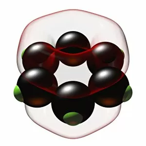
Benzene, molecular model. Benzene consists of a ring of six carbon atoms (black), each with an attached hydrogen atom (green). It is a highly flammable colourless liquid with a characteristic smell

Background imageCompounds Collection: Clenbuterol drug, molecular models

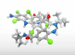
Clenbuterol drug, molecular models, three molecules shown. This decongestant and bronchodilator is used to treat breathing disorders

Background imageCompounds Collection: Bacterial ribosome and protein synthesis

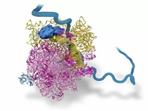
Bacterial ribosome and protein synthesis. Molecular model showing a bacterial ribosome reading an mRNA (messenger ribonucleic acid) strand (blue) and synthesising a protein

Background imageCompounds Collection: Haemagglutinin viral surface protein C015 / 9965

Haemagglutinin viral surface protein C015 / 9965
Haemagglutinin viral surface protein. Molecular model of haemagglutinin, a surface protein from the influenza virus, complexed with a neutralising antibody

Background imageCompounds Collection: Haemagglutinin viral surface protein C015 / 7124

Haemagglutinin viral surface protein C015 / 7124
Haemagglutinin viral surface protein. Molecular model of haemagglutinin, a surface protein from the influenza virus, complexed with a neutralising antibody

Background imageCompounds Collection: Haemagglutinin viral surface protein C015 / 9974

Haemagglutinin viral surface protein C015 / 9974
Haemagglutinin viral surface protein. Molecular model of haemagglutinin, a surface protein from the influenza virus, complexed with a neutralising antibody

Background imageCompounds Collection: Haemagglutinin viral surface protein C015 / 7123

Haemagglutinin viral surface protein C015 / 7123
Haemagglutinin viral surface protein. Molecular model of haemagglutinin, a surface protein from the influenza virus, complexed with a neutralising antibody

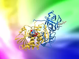
Background imageCompounds Collection: Ricin molecule, artwork C017 / 3656

Ricin molecule, artwork C017 / 3656
Ricin molecule. Computer artwork showing the structure of a molecule of the toxic protein ricin. Ricin comprises two entwined amino acid chains; A (yellow) and B (blue)

Background imageCompounds Collection: Water molecules, artwork C017 / 7384

Water molecules, artwork C017 / 7384
Water molecules. Computer artwork showing the molecular (top) and atomic (bottom) structure of water (H2O). Atoms are colour-coded: hydrogen (blue) and oxygen (white)

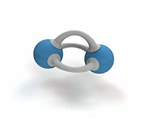
Background imageCompounds Collection: Methane molecule, artwork C017 / 3614

Methane molecule, artwork C017 / 3614
Methane molecule. Computer artwork showing the structure of a molecule of nitrogen (N2). Atoms are colour coded: nitrogen (blue), with the bonds between them as bars (grey)

Background imageCompounds Collection: Ricin molecule, artwork C017 / 3655

Ricin molecule, artwork C017 / 3655
Ricin molecule. Computer artwork showing the structure of a molecule of the toxic protein ricin. Ricin comprises two entwined amino acid chains; A (yellow) and B (blue)

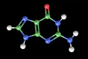
Background imageCompounds Collection: Guanine molecule, artwork C017 / 7239

Guanine molecule, artwork C017 / 7239
Guanine molecule. Computer artwork showing the structure of a molecule of the nucleobase guanine. Atoms are shown as colour-coded spheres: carbon (green), hydrogen (white)

Background imageCompounds Collection: Ricin molecule, artwork C017 / 3648

Ricin molecule, artwork C017 / 3648
Ricin molecule Computer artwork showing the structure of a molecule of the toxic protein ricin (blue and yellow) with an active ribosome in the background

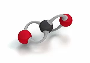
Background imageCompounds Collection: Carbon dioxide molecule C017 / 3600

Carbon dioxide molecule C017 / 3600
Carbon dioxide molecule. Computer artwork showing the structure of a molecule of carbon dioxide (CO2). Atoms are colour coded: oxygen (red) and carbon (black)

Background imageCompounds Collection: Thymine-adenine interaction, artwork C017 / 7368

Thymine-adenine interaction, artwork C017 / 7368
Thymine-adenine interaction. Computer artwork showing the structure of bound thymine and adenine molecules. Atoms are shown as colour-coded spheres: carbon (green), hydrogen (white)

Background imageCompounds Collection: Resveratrol molecule C014 / 2110

Resveratrol molecule C014 / 2110
Resveratrol, molecular model. Resveratrol is a phytoalexin, a defence chemical produced by plants, found in grapes, peanuts, blueberries and some pines

Background imageCompounds Collection: Cytosine molecule, artwork C017 / 7213

Cytosine molecule, artwork C017 / 7213
Cytosine molecule. Computer artwork showing the structure of a molecule of the nucleobase cytosine (2-oxy-4-aminopyrimidine)


All Professionally Made to Order for Quick Shipping

"Exploring the Fascinating World of Compounds: From Copper and Magnesium Sulphate to Graphene" Delving into the intricate beauty of compounds, we witness the mesmerizing sight of copper and magnesium sulphate crystals under a light microscope (LM). A closer look at caffeine crystals through a light micrograph reveals their captivating structure, resembling tiny jewels that fuel our mornings. Oxytocin hormone crystals, captured using polarized light microscopy (PLM C016 / 7196), unveil the remarkable complexity behind this molecule responsible for human bonding. Through an artistic representation, we unravel the secondary structure of proteins – nature's building blocks that orchestrate countless biological processes within us. The perovskite crystal structure captivates scientists with its potential applications in renewable energy technologies, promising a brighter future for sustainable power generation. Another glimpse into oxytocin's world showcases its crystalline form under a light microscope, reminding us of its vital role in nurturing social connections and maternal instincts. Zooming in on caffeine's molecular composition unveils its drug-like qualities that stimulate our nervous system and keep us awake during long nights or early mornings. Peering into the microscopic realm reveals bacterial ribosomes - miniature protein factories essential for life itself - showcasing nature's incredible machinery at work. Cortisol crystals come to life as they are illuminated by a beam of light under a microscope, offering insight into this stress hormone's unique properties within our bodies. Exploring vitamin B12 through its molecular model highlights how this crucial nutrient supports various bodily functions while displaying an elegant arrangement of atoms and bonds. Once again, copper sulphate crystals enchant us with their vibrant colors when observed using a light microscope (LM), reminding us of their diverse industrial uses and chemical significance. Stepping into the realm of materials science brings forth graphene.