Framed Print > Science > SEM
Framed Print : Golgi apparatus, SEM


![]()
Framed Photos From Science Photo Library
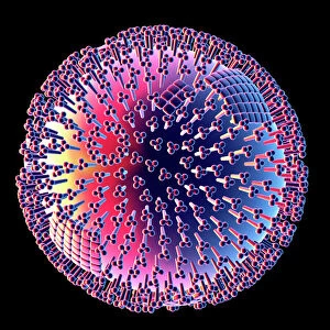
Golgi apparatus, SEM
Golgi apparatus, coloured scanning electron micrograph (SEM). Section through a liver cell showing its Golgi apparatus (grey), a membrane-bound organelle that modifies and packages proteins. Also seen is part of the cell nucleus (orange), which contains the cells genetic information
Science Photo Library features Science and Medical images including photos and illustrations
Media ID 6305923
© DR DAVID FURNESS, KEELE UNIVERSITY/SCIENCE PHOTO LIBRARY
Bodies Cell Biology Crista Cristae Cytology Golgi Apparatus Golgi Body Hepatic Liver Cell Membrane Bound Nucleus Organelle Organelles Vesicle Vesicles False Coloured Section Sectioned
12"x10" Modern Frame
Bring the wonders of science into your home or office with Media Storehouse's Framed Prints featuring the stunning image of the Golgi Apparatus in a coloured Scanning Electron Micrograph (SEM) by Science Photo Library. This captivating print showcases the intricate details of this essential organelle as it modifies and packages proteins in a liver cell. A perfect addition to any space inspiring curiosity and appreciation for the intricacies of life.
10x8 Print in an MDF Wooden Frame with 180 gsm Satin Finish Paper. Glazed using shatter proof thin plexi glass. Frame thickness is 1 inch and depth 0.75 inch. Fluted cardboard backing held with clips. Supplied ready to hang with sawtooth hanger and rubber bumpers. Spot clean with a damp cloth. Packaged foam wrapped in a card.
Contemporary Framed and Mounted Prints - Professionally Made and Ready to Hang
Estimated Image Size (if not cropped) is 25.4cm x 25.4cm (10" x 10")
Estimated Product Size is 30.5cm x 25.4cm (12" x 10")
These are individually made so all sizes are approximate
Artwork printed orientated as per the preview above, with landscape (horizontal) or portrait (vertical) orientation to match the source image.
EDITORS COMMENTS
This print from Science Photo Library showcases the intricate beauty of a liver cell's Golgi apparatus. The coloured scanning electron micrograph (SEM) reveals a section through the cell, highlighting the grey Golgi apparatus, a vital organelle responsible for modifying and packaging proteins within the cell. The image also offers a glimpse of the orange-hued cell nucleus, which contains crucial genetic information. This SEM capture provides an extraordinary level of detail, allowing us to appreciate the complexity and organization within this tiny cellular world. The false-coloured representation adds an artistic touch to this scientific marvel, enhancing our understanding of its structure and function. It is truly remarkable how these microscopic components work together seamlessly to ensure proper protein processing and transportation throughout the liver cell. As we delve into cytology and explore cellular biology further, images like these serve as powerful reminders of nature's incredible design at even its smallest scale. Such visual representations not only inspire awe but also deepen our appreciation for the intricacies that underlie life itself. Science Photo Library continues to provide invaluable resources like this photograph that bridge science with artistry, enabling us to explore new frontiers in biological research while simultaneously celebrating its inherent beauty.
MADE IN THE USA
Safe Shipping with 30 Day Money Back Guarantee
FREE PERSONALISATION*
We are proud to offer a range of customisation features including Personalised Captions, Color Filters and Picture Zoom Tools
SECURE PAYMENTS
We happily accept a wide range of payment options so you can pay for the things you need in the way that is most convenient for you
* Options may vary by product and licensing agreement. Zoomed Pictures can be adjusted in the Basket.