Fraud Blocker Skip to main content

Organelle Collection

Organelles: The Tiny Powerhouses Within Our Cells The world of cells is a fascinating one, filled with intricate structures known as organelles

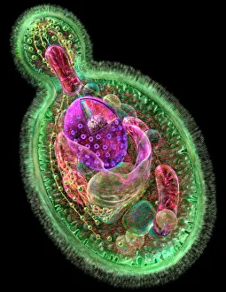
Background imageOrganelle Collection: Budding yeast cell

Budding yeast cell. Computer artwork of asection through a yeast ( Candida albicans )cell that is reproducing asexually. A daughtercell (top left) is budding from the parent cell
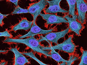
Background imageOrganelle Collection: HeLa cells, light micrograph C017 / 8299

HeLa cells, light micrograph C017 / 8299
HeLa cells, multiphoton fluorescence micrograph (MFM). The cell nuclei, which contain the cells genetic information, are purple. Microtubules are blue and actin microfilaments are red

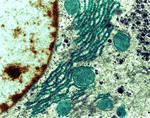
Background imageOrganelle Collection: Rough endoplasmic reticulum, TEM

Rough endoplasmic reticulum, TEM
Rough endoplasmic reticulum, coloured transmission electron micrograph (TEM). This section shows the rough endoplasmic reticulum (ER, folds, centre), a membranous structure that occurs in cells

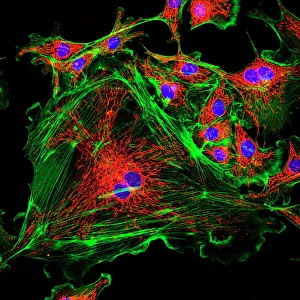
Background imageOrganelle Collection: Cell structure

Cell structure. Confocal light micrograph of cultured endothelial cells. A fluorescent dye has been used to show the cell structure

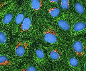
Background imageOrganelle Collection: HeLa cells, light micrograph C017 / 8298

HeLa cells, light micrograph C017 / 8298
HeLa cells, multiphoton fluorescence micrograph (MFM). The cell nuclei, which contain the cells genetic information, are blue. Golgi bodies, which modify and package proteins, are orange

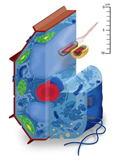
Background imageOrganelle Collection: Cell types, artwork

Cell types, artwork
Cell types. Cutaway artwork with a scale bar (upper right, in micrometres) showing the relative sizes of eukaryotic cells (those containing a nucleus) and prokaryotic cells (those lacking a nucleus)

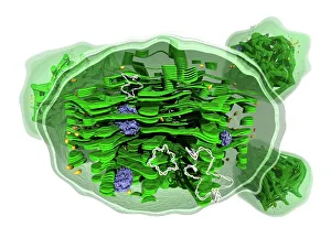
Background imageOrganelle Collection: Chloroplast structure, artwork

Chloroplast structure, artwork
Chloroplast structure. Artwork showing the internal structure of chloroplasts, the organelles in plant cells responsible for photosynthesis

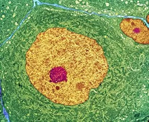
Background imageOrganelle Collection: Nerve cell, TEM

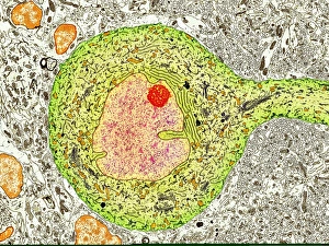
Nerve cell, TEM
Nerve cell. Coloured transmission electron micrograph (TEM) of a nerve cell body in cross- section. The cell has a large nucleus (yellow) and inner nucleolus (red)

Background imageOrganelle Collection: Mitochondrion, TEM

Mitochondrion, TEM
Mitochondrion. Coloured transmission electron micrograph (TEM) of mitochondria (green/blue) in heart muscle. Mitochondria are organelles found in the cytoplasm of eukaryotic cells

Background imageOrganelle Collection: Fibroblast cell, artwork

Fibroblast cell, artwork
Fibroblast cell. Computer artwork of a fibroblast excreting collagen fibres (tropocollagen). Fibroblasts are cells that produce connective tissue such as collagen (tropocollagen)

Background imageOrganelle Collection: Golgi apparatus, SEM

Golgi apparatus, SEM
Golgi apparatus, coloured scanning electron micrograph (SEM). Section through a liver cell showing its Golgi apparatus (grey), a membrane-bound organelle that modifies and packages proteins

Background imageOrganelle Collection: Purkinje nerve cell, TEM C014 / 0583

Purkinje nerve cell, TEM C014 / 0583
Purkinje nerve cell. Transmission electron micrograph (TEM) of a purkinje nerve cell (green) from the cerebellum of the brain, showing the cell body (centre) and its primary dendrite (cell process)

Background imageOrganelle Collection: Animal cell structure

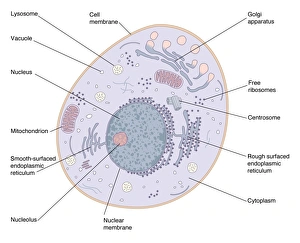
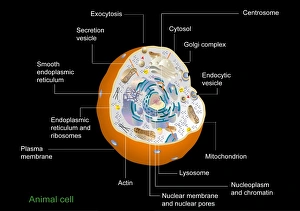
Animal cell structure. Artwork of a section through an animal cell. At lower centre is the nucleus, which contains the cells genetic information in the form of DNA (deoxyribonucleic acid)

Background imageOrganelle Collection: Microscopic view of animal cell

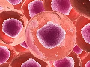
Microscopic view of animal cell

Background imageOrganelle Collection: Animal cell anatomy, diagram

Animal cell anatomy, diagram
Animal cell anatomy. Diagram showing the internal and external anatomy of an animal cell

Background imageOrganelle Collection: Chloroplast, SEM

Chloroplast, SEM
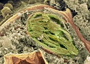
Chloroplast. Coloured scanning electron micrograph (SEM) of a section through a plant cell, showing a fractured chloroplast (green)

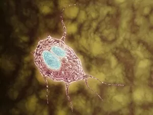
Background imageOrganelle Collection: Microscopic view of Giardiasis

Microscopic view of Giardiasis, an infectious disease caused by a unicellular parasite known as Giardia lamblia

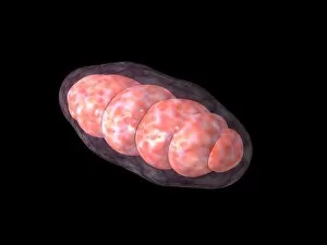
Background imageOrganelle Collection: Conceptual image of mitochondria

Conceptual image of mitochondria

Background imageOrganelle Collection: Pancreatic acinar cell

Pancreatic acinar cell. Transmission electron micrograph (TEM) of a section through an enzyme-secreting acinar cell in the human pancreas, showing part of the nucleus (round, far left)

Background imageOrganelle Collection: Hepatocyte liver cell, TEM

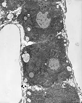
Hepatocyte liver cell, TEM
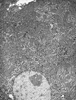
Hepatocyte liver cell. Transmission electron micrograph (TEM) of a section through an hepatocyte liver cell, showing rough and smooth endoplasmic reticulum (ER, dark lines)

Background imageOrganelle Collection: Liver tissue, TEM

Liver tissue, TEM
Liver tissue. Transmission electron micrograph (TEM) of a section through the liver, showing part of a radial cord of hepatocyte liver cells (dark) and the vascular sinusoids (white)

Background imageOrganelle Collection: Golgi apparatus, TEM C017 / 8234

Golgi apparatus, TEM C017 / 8234
Golgi apparatus. Transmission electron micrograph (TEM) of Golgi apparatus from a Chlamydomonas sp. alga. Golgi are membrane-bound organelles that modify and package proteins

Background imageOrganelle Collection: Skeletal muscle, TEM C016 / 5369

Skeletal muscle, TEM C016 / 5369
Skeletal muscle. Transmission electron micrograph (TEM) of a cross section through human skeletal (striated) muscle. Blocks of muscle (lighter grey) are surrounded by connective tissue (black)

Background imageOrganelle Collection: Skin cancer cell, SEM C016 / 4764

Skin cancer cell, SEM C016 / 4764
Skin cancer cell. Coloured ion-abrasion scanning electron micrograph (IA-SEM) of a melanoma cell. The large dark sphere is the cell nucleus, which contains the cells genetic information

Background imageOrganelle Collection: Cardiac muscle, TEM

Cardiac muscle, TEM
Cardiac muscle. Coloured transmission electron micrograph (TEM) of cardiac muscle fibrils (orange)from a healthy heart. Mitochondria (pink) supply the muscle cells with energy

Background imageOrganelle Collection: Cardiac muscle and capillary, TEM

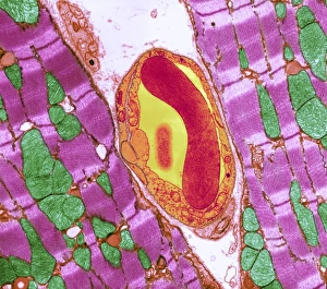
Cardiac muscle and capillary, TEM
Cardiac muscle. Coloured transmission electron micrograph (TEM) of cardiac muscle fibrils (purple)from a healthy heart. Mitochondria (green) supply the muscle cells with energy

Background imageOrganelle Collection: Mitochondria, TEM

Mitochondria, TEM
Mitochondria. Coloured transmission electron micrograph (TEM) of mitochondria (red) in heart muscle. Mitochondria are organelles found in the cytoplasm of eukaryotic cells

Background imageOrganelle Collection: Mitochondria, SEM

Mitochondria, SEM
Mitochondria. Coloured scanning electron micrograph (SEM) of mitochondria (red) in a kidney cell. Mitochondria are a type of organelle found in the cytoplasm of eukaryotic cells

Background imageOrganelle Collection: Illustration of how an amoeba moves, liquid cytoplasm flowing through pseudopods carrying

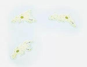
Illustration of how an amoeba moves, liquid cytoplasm flowing through pseudopods carrying organelles with it, amoeba sending out pseudopods in direction of motion

Background imageOrganelle Collection: Microscopic view inside of the artery with intestinal villi

Microscopic view inside of the artery with intestinal villi

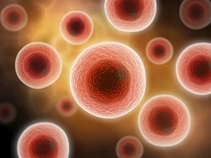
Background imageOrganelle Collection: Microscopic view of cell

Microscopic view of cell

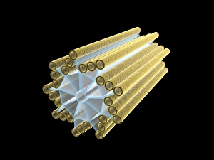
Background imageOrganelle Collection: Conceptual image of centriole

Conceptual image of centriole

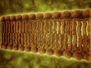
Background imageOrganelle Collection: Microscopic view of phospholipids

Microscopic view of phospholipids. Phospholipids are a major component of all cell membranes as they can form lipid bilayers

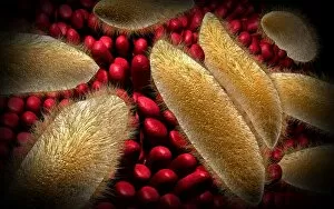
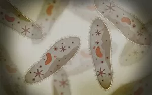
Background imageOrganelle Collection: Conceptual image of paramecium

Conceptual image of paramecium

Background imageOrganelle Collection: Conceptual image of chloroplast

Conceptual image of chloroplast

Background imageOrganelle Collection: Conceptual image of intestinal villi

Conceptual image of intestinal villi

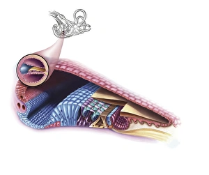
Background imageOrganelle Collection: Interior detail of the cochlea

Interior detail of the cochlea

Background imageOrganelle Collection: Cell nucleus with chromosome

Cell nucleus with chromosome. The cell nucleus helps control eating, movement, and reproduction

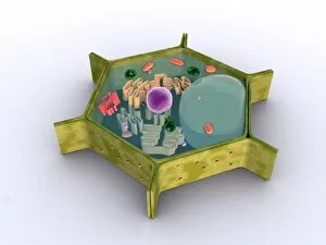
Background imageOrganelle Collection: Conceptual image of a plant cell and its components

Conceptual image of a plant cell and its components

Background imageOrganelle Collection: Microscopic view of paramecium

Microscopic view of paramecium

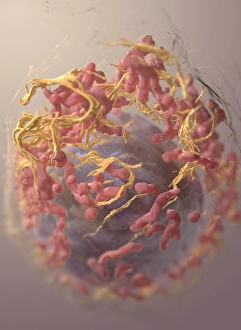
Background imageOrganelle Collection: 3D structure of melanoma cell

3D structure of melanoma cell
3D structure of a melanoma cell derived by ion abrasion scanning electron microscopy

Background imageOrganelle Collection: Conceptual image of human cell

Conceptual image of human cell

Background imageOrganelle Collection: Human cell anatomy

Human cell anatomy

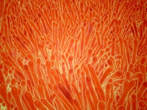
Background imageOrganelle Collection: Microscopic view of intestinal villi inside the small intestine

Microscopic view of intestinal villi inside the small intestine
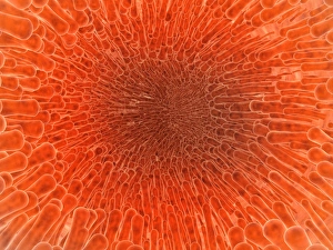
Microscopic view of intestinal villi which can be found inside of the small intestine

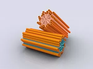
Background imageOrganelle Collection: Conceptual image of centrioles

Conceptual image of centrioles

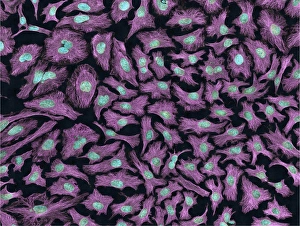
Background imageOrganelle Collection: Multiphoton fluorescence image of HeLa cells

Multiphoton fluorescence image of HeLa cells with cytoskeletal microtubules (magenta) and DNA (cyan)

Background imageOrganelle Collection: Conceptual image of human cytomegalovirus

Conceptual image of human cytomegalovirus. Cytomegalovirus is a genus of the viral family Herpesviridae

Background imageOrganelle Collection: Microscopic view of Mitochondria

Microscopic view of Mitochondria. Mitochondria are the cells power producers. They convert energy into forms that are usable by the cell. Located in the cytoplasmcells activities


All Professionally Made to Order for Quick Shipping

Organelles: The Tiny Powerhouses Within Our Cells The world of cells is a fascinating one, filled with intricate structures known as organelles. These tiny entities play crucial roles in maintaining the functionality and survival of our cells. From budding yeast to HeLa cells, each cell type possesses its unique set of organelles that contribute to their specialized functions. Under the lens of a light microscope, the HeLa cells reveal mesmerizing patterns resembling abstract artwork. Their delicate structures are visible, showcasing the complexity within these microscopic powerhouses. Meanwhile, another image captures nerve cells in all their glory - long and slender extensions reaching out like branches from a tree. Zooming in further using transmission electron microscopy (TEM), we get an up-close look at some specific organelles. The rough endoplasmic reticulum appears as a network of interconnected membranes studded with ribosomes responsible for protein synthesis. Mitochondria steal the spotlight next; these bean-shaped powerhouses generate energy for cellular activities through respiration. But it's not just animal cells that possess remarkable organelles; plant cells have their own unique features too. Chloroplasts, depicted beautifully through artwork, capture sunlight and convert it into energy via photosynthesis—a process vital for sustaining life on Earth. Another TEM image reveals Purkinje nerve cells—large neurons found in our brain's cerebellum—showcasing their intricate branching structure responsible for coordinating movement and balance. Finally, scanning electron microscopy (SEM) unveils the Golgi apparatus—an organelle involved in processing proteins and packaging them into vesicles for transportation throughout the cell or secretion outside it. These glimpses into different types of organelles highlight their diverse forms and functions within our cellular world. They remind us that even though they may be invisible to the naked eye, these minuscule entities hold immense importance in keeping our bodies functioning harmoniously at every level.