Fraud Blocker Skip to main content

Vesicles Collection

Vesicles: Tiny Transporters in the Intricate World of Cells In the fascinating realm of cellular biology, vesicles play a crucial role as miniature transporters

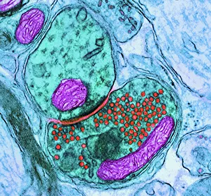
Background imageVesicles Collection: Synapse nerve junction, TEM

Synapse nerve junction, TEM
Synapse. Coloured transmission electron micrograph (TEM) of a synapse, a junction between two nerve cells, in the brain. At a synapse an electrical signal is transmitted from one cell to the next in

Background imageVesicles Collection: Rough endoplasmic reticulum, TEM

Rough endoplasmic reticulum, TEM
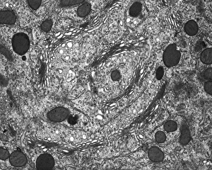
Rough endoplasmic reticulum, coloured transmission electron micrograph (TEM). This section shows the rough endoplasmic reticulum (ER, folds, centre), a membranous structure that occurs in cells

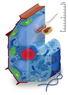
Background imageVesicles Collection: Cell types, artwork

Cell types, artwork
Cell types. Cutaway artwork with a scale bar (upper right, in micrometres) showing the relative sizes of eukaryotic cells (those containing a nucleus) and prokaryotic cells (those lacking a nucleus)

Background imageVesicles Collection: Fibroblast cell, artwork

Fibroblast cell, artwork
Fibroblast cell. Computer artwork of a fibroblast excreting collagen fibres (tropocollagen). Fibroblasts are cells that produce connective tissue such as collagen (tropocollagen)

Background imageVesicles Collection: Golgi apparatus, SEM

Golgi apparatus, SEM
Golgi apparatus, coloured scanning electron micrograph (SEM). Section through a liver cell showing its Golgi apparatus (grey), a membrane-bound organelle that modifies and packages proteins

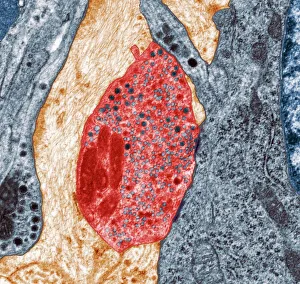
Background imageVesicles Collection: Nerve synapse, TEM

Nerve synapse, TEM
Nerve synapse. Coloured transmission electron micrograph (TEM) of the neuron (nerve) terminal at a synapse in the diaphragm

Background imageVesicles Collection: Nasal lining, SEM

Nasal lining, SEM
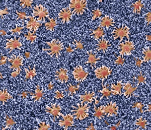
Nasal lining. Coloured scanning electron micrograph (SEM) of the olfactory epithelium that lines the nasal cavity, showing olfactory cells (red) surrounded by numerous cilia (hair-like projections)

Background imageVesicles Collection: Shingles nerve damage

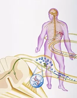
Shingles nerve damage

Background imageVesicles Collection: Model of animal cell, including cell nucleus, golgi body, lysosomes, centrioles, mitochondria

Model of animal cell, including cell nucleus, golgi body, lysosomes, centrioles, mitochondria, endoplasmic reticulum, ribosomes, cytoplasm, vesicles, thin plasma membrane

Background imageVesicles Collection: Bladder lining, TEM C014 / 1473

Bladder lining, TEM C014 / 1473
Bladder lining. Transmission electron micrograph (TEM) of a section through the epithelial cells that line of a human bladder, showing large flat vesicles (large, blue)

Background imageVesicles Collection: Stem cell dying, SEM

Stem cell dying, SEM
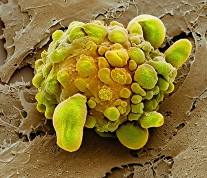
Stem cell dying. Coloured scanning electron micrograph (SEM) of a stem cell undergoing apoptosis, or programmed cell death. Apoptosis occurs when a cell becomes old or damaged

Background imageVesicles Collection: Smell receptor, TEM

Smell receptor, TEM
Smell receptor. Transmission electron micrograph (TEM) of a section through the olfactory epithelium that lines the nasal cavity, showing an olfactory cell (smell receptor)

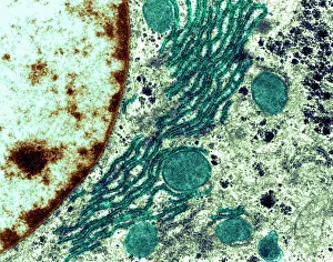
Background imageVesicles Collection: Golgi membranes, TEM

Golgi membranes, TEM
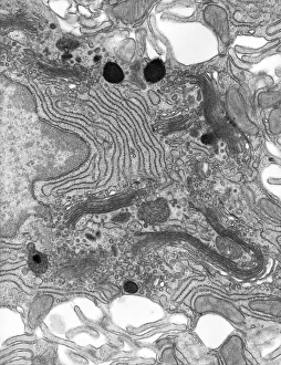
Golgi membranes. Transmission electron micrograph (TEM) of a section through a cell, showing the membranes (dark lines) of the Golgi apparatus

Background imageVesicles Collection: Enterocyte, TEM

Enterocyte, TEM
Enterocyte. Transmission electron micrograph (TEM) of a section through the cytoplasm and part of the nucleus of an enterocyte cell located in a crypt of Lieberkuhn of the small intestine

Background imageVesicles Collection: Clathrin lattice, molecular model C018 / 0453

Clathrin lattice, molecular model C018 / 0453
Clathrin lattice, molecular model. This polyhedral protein lattice coats eukaryotic cell membranes (vesicles) and is involved in protein secretion and membrane trafficking

Background imageVesicles Collection: Clathrin lattice, molecular model C018 / 0452

Clathrin lattice, molecular model C018 / 0452
Clathrin lattice, molecular model. This polyhedral protein lattice coats eukaryotic cell membranes (vesicles) and is involved in protein secretion and membrane trafficking

Background imageVesicles Collection: Clathrin lattice, molecular model C018 / 0454

Clathrin lattice, molecular model C018 / 0454
Clathrin lattice, molecular model. This polyhedral protein lattice coats eukaryotic cell membranes (vesicles) and is involved in protein secretion and membrane trafficking

Background imageVesicles Collection: Intestinal arteriole, TEM

Intestinal arteriole, TEM
Intestinal arteriole. Transmission electron micrograph (TEM) of a section through an arteriole in the wall of the small intestine. Magnification: x5000 when printed 10 centimetres wide

Background imageVesicles Collection: Muscle arteriole, TEM

Muscle arteriole, TEM
Muscle arteriole. Transmission electron micrograph (TEM) of a section through an arteriole (black, centre) in striated muscle tissue. Magnification: x3500 when printed 10 centimetres wide

Background imageVesicles Collection: Clathrin lattice, molecular model C015 / 6788

Clathrin lattice, molecular model C015 / 6788
Molecular model of a complete clathrin lattice. The polyhedral protein lattice coats eukaryotic cell membranes (vesicles) and coated pits and appears to be involved in protein secretion

Background imageVesicles Collection: Clathrin lattice, molecular model C015 / 6790

Clathrin lattice, molecular model C015 / 6790
Molecular model of a complete clathrin lattice. The polyhedral protein lattice coats eukaryotic cell membranes (vesicles) and coated pits and appears to be involved in protein secretion

Background imageVesicles Collection: Clathrin lattice, molecular model C015 / 6770

Clathrin lattice, molecular model C015 / 6770
Molecular model of a complete clathrin lattice. The polyhedral protein lattice coats eukaryotic cell membranes (vesicles) and coated pits and appears to be involved in protein secretion

Background imageVesicles Collection: HeLa cell dying, SEM C017 / 8304

HeLa cell dying, SEM C017 / 8304
HeLa cell dying, coloured scanning electron micrograph (SEM). The cell in the foreground is undergoing apoptosis, or programmed cell death. Apoptosis occurs when a cell becomes old or damaged

Background imageVesicles Collection: Intracellular transport, artwork C013 / 4997

Intracellular transport, artwork C013 / 4997
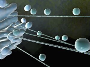
Intracellular transport. Computer artwork of vesicles (spheres) being transported from a Golgi body (blue, left) around the cell by microtubules (string-like)

Background imageVesicles Collection: Intracellular transport, artwork C013 / 4995

Intracellular transport, artwork C013 / 4995
Intracellular transport. Computer artwork of vesicles (spheres) being transported from a Golgi body (blue, top) around the cell by microtubules (string-like)

Background imageVesicles Collection: Pancreas cells, SEM

Pancreas cells, SEM
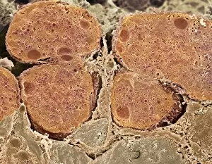
Pancreas cells. Coloured scanning electron micrograph (SEM) of acinar (exocrine) pancreatic cells. Acinar cells produce and excrete digestive enzymes to the small intestine, via the pancreatic ducts

Background imageVesicles Collection: Alveolar cell, TEM

Alveolar cell, TEM
Alveolar cell. Coloured transmission electron micrograph (TEM) of a section through a type II alveolar cell. This cell is found in the alveoli (air sacs) of the lungs

Background imageVesicles Collection: Neuromuscular synapse, light micrograph

Neuromuscular synapse, light micrograph
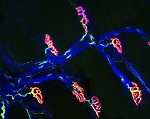
Neuromuscular junction. Fluorescent confocal light micrograph of the junction between a nerve cell and a muscle (not seen). The axon of the nerve cell (neuron) has been tagged with a blue dye

Background imageVesicles Collection: Coloured TEM of a nerve synapse

Coloured TEM of a nerve synapse

Background imageVesicles Collection: Astrocyte / neuron synapse in the brain

Astrocyte / neuron synapse in the brain
Astrocyte/neuron synapse in the brain. Artwork of a nerve cell (neuron, yellow) synapse in the brain with an associated regulatory astrocyte cell (red)

Background imageVesicles Collection: Shingles

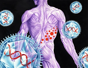
Shingles. Artwork of varicella-zoster virus particles (virions, blue) during shingles. Each viron comprises an icosahedral (20-sided) capsid containing viral DNA

Background imageVesicles Collection: Skin lesions

Skin lesions. Artwork from Robert Willans 1808 medical textbook, On Cutaneous Diseases, showing 16 different kinds of disorders affecting the skin

Background imageVesicles Collection: Simulated space ice

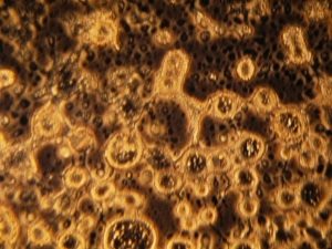
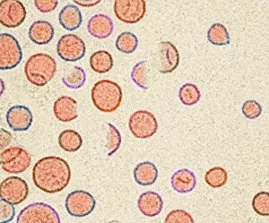
Simulated space ice. Light micrograph of vesicles (round structures) in ice formed under conditions simulating those in space

Background imageVesicles Collection: Liposomes, SEM

Liposomes, SEM

Background imageVesicles Collection: Liposomes, TEM

Liposomes, TEM

Background imageVesicles Collection: Pancreas cell, SEM

Pancreas cell, SEM
Pancreas cell. Coloured scanning electron micrograph (SEM) of an acinar (exocrine) pancreatic cell. Acinar cells produce and excrete digestive enzymes to the small intestine

Background imageVesicles Collection: Basophil white blood cells, artwork

Basophil white blood cells, artwork
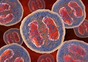
Basophil white blood cells. Computer artwork of basophil white blood cells, showing their lobed nuclei (red). Basophils are the smallest and least common of the white blood cells

Background imageVesicles Collection: Cytokinesis, artwork

Cytokinesis, artwork
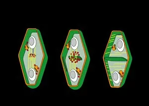
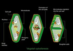
Cytokinesis. Artwork showing the stage of cell division that involves the splitting of the cell cytoplasm between two daughter cells

Background imageVesicles Collection: Cytokinesis, diagram

Cytokinesis, diagram
Cytokinesis. Diagram showing the stage of cell division that involves the splitting of the cell cytoplasm between two daughter cells

Background imageVesicles Collection: Familial hypercholesterolaemia, diagram

Familial hypercholesterolaemia, diagram
Familial hypercholesterolaemia. Diagram showing the biochemical processes that take place in a case of the genetic disorder familial hypercholesterolaemia

Background imageVesicles Collection: Meningitis B vesicles, TEM

Meningitis B vesicles, TEM
Meningitis B vesicles. Coloured transmission electron micrograph (TEM) of outer membrane vesicles (OMV) from Neisseria meningitidis serogroup B bacteria

Background imageVesicles Collection: Cell structure, fluorescent micrograph

Cell structure, fluorescent micrograph
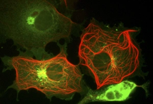
Cell structure. Fluorescent light micrograph of cultured cells from a cell line derived from African green monkey kidney cells. Microtubules, part of the cells cytoskeleton, are red

Background imageVesicles Collection: Animal cell processes, artwork

Animal cell processes, artwork
Animal cell processes. Cutaway artwork showing the structures inside an animal cell and four different processes that take place inside it or on its membrane (all marked by magnifying glasses)

Background imageVesicles Collection: Animal cell structure, artwork

Animal cell structure, artwork
Animal cell structure. Artwork showing the internal structure of an animal cell. Inside the cell, the cellular structures (organelles) include the Golgi apparatus (green)

Background imageVesicles Collection: Golgi apparatus, artwork

Golgi apparatus, artwork. This structure is an organelle found within eukaryotic cells. It receives proteins and lipids that are synthesised elsewhere on the endoplasmic reticulum

Background imageVesicles Collection: Protein, microtubules and cell, artwork

Protein, microtubules and cell, artwork
3D computer artwork of a protein attached to microtubules, transporting vesicles and other important parts of the cell. Microtubules are polymers of the protein tubulin
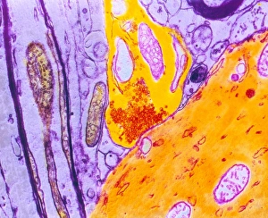
Background imageVesicles Collection: Axon anatomy, diagram

Axon anatomy, diagram
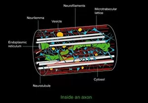
Axon anatomy. Diagram of the anatomical structure of an axon, the main extension (dendrite) of a nerve cell. Cellular components include endoplasmic reticulum (green), neurotubules (white)


All Professionally Made to Order for Quick Shipping

Vesicles: Tiny Transporters in the Intricate World of Cells In the fascinating realm of cellular biology, vesicles play a crucial role as miniature transporters, shuttling vital substances within cells and ensuring their proper functioning. These tiny sacs are involved in various processes, from synapse nerve junctions to protein synthesis. Under the powerful lens of a transmission electron microscope (TEM), we can observe vesicles at work. At synapse nerve junctions, these dynamic structures facilitate communication between neurons by transmitting chemical signals. The TEM reveals their intricate network, highlighting their significance in maintaining healthy neural connections. Another intriguing sight captured by TEM is the rough endoplasmic reticulum (ER). This labyrinthine structure studded with ribosomes produces proteins destined for export or insertion into cell membranes. Vesicles bud off from this ER network, carrying newly synthesized proteins to different parts of the cell. Cell types come alive through captivating artwork depicting fibroblast cells and protein interactions with microtubules. These illustrations showcase how vesicles collaborate with other cellular components to ensure proper functioning and organization within cells. The Golgi apparatus takes center stage under scanning electron microscopy (SEM). Its distinctive appearance resembles a stack of flattened pancakes and serves as an important processing center for proteins before they are packaged into vesicles for transportation throughout the cell. Nasal lining viewed through SEM provides insight into its complex structure and highlights how vesicles contribute to maintaining respiratory health. Additionally, shingles nerve damage reminds us that disruptions in vesicle transport can lead to severe consequences such as pain and discomfort. A detailed model of an animal cell offers a comprehensive view of various organelles including nuclei, golgi bodies, lysosomes, centrioles, mitochondria - all interconnected by a vast network of endoplasmic reticulum. Vesicles stand out among these essential components as key players responsible for transporting molecules across different compartments within the cell.