Fine Art Print : MRI brain scan and nerve cells F006 / 3595

![]()
Fine Art Prints From Science Photo Library
MRI brain scan and nerve cells F006 / 3595
Computer artwork of a sagittal (side) view of a human head. The largest part of the brain is the cerebrum (upper part). This surrounds the limbic system (curved structures at upper centre). Below this, the brainstem runs downwards to become the spinal cord. To the right of the brainstem is the cerebellum (branched structure). The blocks of the vertebrae in the neck are seen to the left of the spinal cord. In the background a neural network of nerve cells firing
Science Photo Library features Science and Medical images including photos and illustrations
Media ID 9333829
© PASIEKA/SCIENCE PHOTO LIBRARY
Activity Brain Scan Brain Stem Central Nervous System Cerebellum Cerebrum Firing Intelligence Nerve Network Neural Neural Network Sagittal Scan Spinal Chord Brain Cells Internal Organ Nervous System Neurological Neurology
20"x16" (+3" Border) Fine Art Print
Discover the intricacies of the human brain with our Fine Art Print from Media Storehouse and Science Photo Library. Featuring the captivating image F006 / 3595 by PASIEKA/SCIENCE PHOTO LIBRARY, this print showcases a computer-generated, sagittal view of a human head. The largest part of the brain, the cerebrum, is prominently displayed, encircling the limbic system. This stunning piece is not just a work of art, but an invitation to explore the complexities of the nervous system. Bring this mesmerizing print into your home or office and ignite curiosity and conversation.
20x16 image printed on 26x22 Fine Art Rag Paper with 3" (76mm) white border. Our Fine Art Prints are printed on 300gsm 100% acid free, PH neutral paper with archival properties. This printing method is used by museums and art collections to exhibit photographs and art reproductions.
Our fine art prints are high-quality prints made using a paper called Photo Rag. This 100% cotton rag fibre paper is known for its exceptional image sharpness, rich colors, and high level of detail, making it a popular choice for professional photographers and artists. Photo rag paper is our clear recommendation for a fine art paper print. If you can afford to spend more on a higher quality paper, then Photo Rag is our clear recommendation for a fine art paper print.
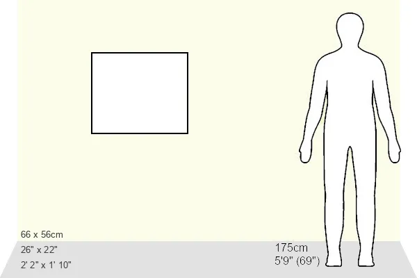
Estimated Image Size (if not cropped) is 50.8cm x 40.6cm (20" x 16")
Estimated Product Size is 66cm x 55.9cm (26" x 22")
These are individually made so all sizes are approximate
Artwork printed orientated as per the preview above, with landscape (horizontal) orientation to match the source image.
EDITORS COMMENTS
This print showcases the intricate beauty of the human brain and its complex network of nerve cells. In this computer artwork, we are presented with a sagittal (side) view of a human head, allowing us to explore the various components that make up this remarkable organ. At first glance, our attention is drawn to the largest part of the brain - the cerebrum - which occupies the upper portion. Surrounding it like a protective embrace is the limbic system, characterized by its elegant curved structures at the upper center. As we shift our gaze downwards, we encounter the brainstem gracefully descending to become the spinal cord. To complement this visual symphony, onlookers can appreciate another vital component situated to the right of the brainstem: The cerebellum. Its branched structure adds an element of complexity and intrigue to this already mesmerizing composition. Intriguingly juxtaposed against a black background, we witness a neural network in action as nerve cells fire with purpose and intensity. This dynamic display represents activity within our nervous system – an essential aspect contributing to intelligence and overall health. Captured through medical illustration techniques by PASIEKA/SCIENCE PHOTO LIBRARY, this image serves as both an artistic masterpiece and scientific marvel. It invites viewers into an awe-inspiring world where artistry meets medicine; where understanding meets wonderment; where science illuminates what lies beneath our skin.
MADE IN THE USA
Safe Shipping with 30 Day Money Back Guarantee
FREE PERSONALISATION*
We are proud to offer a range of customisation features including Personalised Captions, Color Filters and Picture Zoom Tools
SECURE PAYMENTS
We happily accept a wide range of payment options so you can pay for the things you need in the way that is most convenient for you
* Options may vary by product and licensing agreement. Zoomed Pictures can be adjusted in the Basket.