Fine Art Print : Intestinal brush border, TEM

![]()
Fine Art Prints From Science Photo Library
Intestinal brush border, TEM
Intestinal brush border. Transmission electron micrograph (TEM) of intestinal absorptive cells sectioned horizontally at their apex to show the surface microvilli (round). Microvilli are slender finger-like extensions of the intestinal cell that serve to greatly increase the surface area for absorption of nutrients from the lumen of the gastrointestinal tract. The brush borders of the intestinal lining are the site of terminal carbohydrate digestion. The microvilli have enzymes for this final part of digestion anchored into their apical plasma membrane as integral membrane proteins. These enzymes are found near to the transporters that will then allow absorption of the digested nutrients. Magnification: x6, 000 when printed at 10 centimetres wide
Science Photo Library features Science and Medical images including photos and illustrations
Media ID 9240563
© MICROSCAPE/SCIENCE PHOTO LIBRARY
Absorption Black And White Brush Border Cell Surface Colored Digestion Digestive System Epithelial Epithelium Gastroenterological Gastroenterology Gastrointestinal Histological Histology Intestinal Intestine Intestines Lining Microvilli Microvillus Transmission Electron Micrograph Transmission Electron Microscope Transverse Section Villi Villus Cells Section Sectioned
20"x16" (+3" Border) Fine Art Print
Discover the intricacies of the human body with our Fine Art Prints from Media Storehouse. This captivating image, captured through Transmission Electron Microscopy by Science Photo Library, showcases the intestinal brush border in exquisite detail. Witness the intricate surface of absorptive cells, where microvilli form a complex network, enhancing nutrient absorption. Bring the wonders of science into your home or office with our high-quality, museum-grade prints. Each print is carefully crafted to preserve the original image's detail and vibrancy, making for a stunning addition to any space.
20x16 image printed on 26x22 Fine Art Rag Paper with 3" (76mm) white border. Our Fine Art Prints are printed on 300gsm 100% acid free, PH neutral paper with archival properties. This printing method is used by museums and art collections to exhibit photographs and art reproductions.
Our fine art prints are high-quality prints made using a paper called Photo Rag. This 100% cotton rag fibre paper is known for its exceptional image sharpness, rich colors, and high level of detail, making it a popular choice for professional photographers and artists. Photo rag paper is our clear recommendation for a fine art paper print. If you can afford to spend more on a higher quality paper, then Photo Rag is our clear recommendation for a fine art paper print.
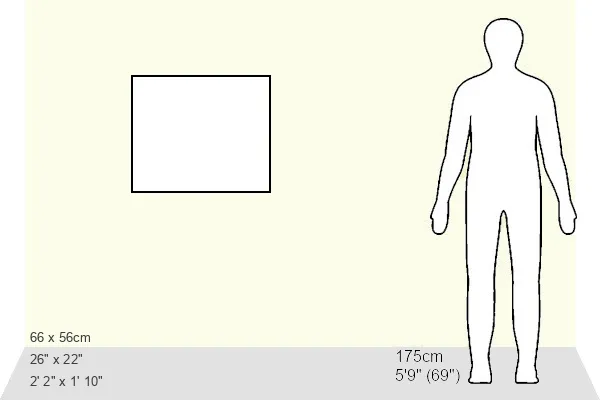
Estimated Image Size (if not cropped) is 50.8cm x 40.6cm (20" x 16")
Estimated Product Size is 66cm x 55.9cm (26" x 22")
These are individually made so all sizes are approximate
Artwork printed orientated as per the preview above, with landscape (horizontal) orientation to match the source image.
EDITORS COMMENTS
This print from Science Photo Library showcases the intricate details of the intestinal brush border, as seen through a transmission electron microscope (TEM). The image reveals a section of horizontally cut absorptive cells in the intestine, specifically highlighting their surface microvilli. These slender finger-like extensions play a crucial role in increasing the surface area for nutrient absorption from the gastrointestinal tract. The brush borders of the intestinal lining serve as the site for terminal carbohydrate digestion. Anchored into their apical plasma membrane, these microvilli house enzymes responsible for this final stage of digestion. Positioned near transporters that facilitate nutrient absorption, these enzymes ensure efficient uptake of digested nutrients. With a magnification level of x6,000 when printed at 10 centimeters wide, this print offers an up-close and detailed view of this vital aspect of our digestive system. The colored and black-and-white composition adds depth to its visual appeal while emphasizing its biological significance. Ideal for biology enthusiasts or those studying gastroenterology, this image provides valuable insights into the structure and function of our gut epithelium. It serves as a reminder that even on a microscopic scale, our body's systems work harmoniously to maintain health and enable proper digestion and absorption processes within us.
MADE IN THE USA
Safe Shipping with 30 Day Money Back Guarantee
FREE PERSONALISATION*
We are proud to offer a range of customisation features including Personalised Captions, Color Filters and Picture Zoom Tools
SECURE PAYMENTS
We happily accept a wide range of payment options so you can pay for the things you need in the way that is most convenient for you
* Options may vary by product and licensing agreement. Zoomed Pictures can be adjusted in the Basket.