Fine Art Print : Brain tumour, DTI MRI scan C017 / 7057

![]()
Fine Art Prints From Science Photo Library
Brain tumour, DTI MRI scan C017 / 7057
Brain tumour. 3D diffusion tensor imaging (DTI) magnetic resonance imaging (MRI) scan of nerve pathways (coloured) in a brain with a tumour (red, centre right). The brain is seen from the side, with the front of the brain at right. Brain tumours can be benign or malignant (cancerous). They can cause seizures, headaches, and memory and personality changes due to the growth of the tumour. The nerve fibres include the corticospinal tract (blue), passing from the motor cortex to the spinal cord. Diffusion tensor imaging (tractography) measures the direction of water diffusion, which in the brain reveals the orientation of nerve fibres
Science Photo Library features Science and Medical images including photos and illustrations
Media ID 9270275
© SHERBROOKE CONNECTIVITY IMAGING LAB/SCIENCE PHOTO LIBRARY
Brain Imaging Brain Scan Cancer Cancerous Central Nervous System Cerebral Diffusion Tensor Imaging Dti Scan Fiber Fibers Fibre Fibres Imaging Technique Magnetic Resonance Imaging Malignancy Malignant Motor Cortex Pathway Mri Scan Mri Scanner Nerve Nerve Fibre Nerves Neural Pathway Neural Tract Neuropathology Oncology Paths Pathway Pathways Structural Tractogram Tractography Tumor Tumour Brain Condition Disorder Neurological Neurology
20"x16" (+3" Border) Fine Art Print
Discover the intricacies of the human brain with our Fine Art Print from Media Storehouse, featuring a captivating 3D diffusion tensor imaging (DTI) MRI scan of nerve pathways in a brain with a tumour, provided by SHERBROOKE CONNECTIVITY IMAGING LAB/SCIENCE PHOTO LIBRARY (C017 / 7057). This stunning image, showcasing the vibrant colours of healthy nerve pathways contrasting against the red tumour, offers a unique perspective into the complexities of neurology. Bring this mesmerizing piece of art into your home or office to inspire curiosity and ignite conversations about the wonders of the human body.
20x16 image printed on 26x22 Fine Art Rag Paper with 3" (76mm) white border. Our Fine Art Prints are printed on 300gsm 100% acid free, PH neutral paper with archival properties. This printing method is used by museums and art collections to exhibit photographs and art reproductions.
Our fine art prints are high-quality prints made using a paper called Photo Rag. This 100% cotton rag fibre paper is known for its exceptional image sharpness, rich colors, and high level of detail, making it a popular choice for professional photographers and artists. Photo rag paper is our clear recommendation for a fine art paper print. If you can afford to spend more on a higher quality paper, then Photo Rag is our clear recommendation for a fine art paper print.
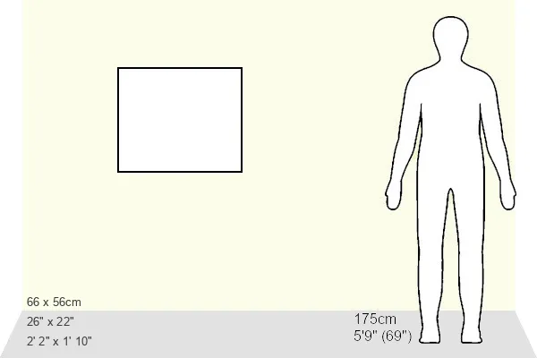
Estimated Image Size (if not cropped) is 48.4cm x 40.6cm (19.1" x 16")
Estimated Product Size is 66cm x 55.9cm (26" x 22")
These are individually made so all sizes are approximate
Artwork printed orientated as per the preview above, with landscape (horizontal) orientation to match the source image.
EDITORS COMMENTS
This print showcases a 3D diffusion tensor imaging (DTI) magnetic resonance imaging (MRI) scan of a brain with a prominent red tumor at its center-right. The nerve pathways within the brain are color-coded, providing an intricate visual representation of the neural connections affected by this condition. From the side view, we can observe the front of the brain on the right. Brain tumors can be either benign or malignant, and their growth often leads to various symptoms such as seizures, headaches, and changes in memory and personality. This image highlights how these tumors disrupt normal brain function by encroaching upon vital nerve fibers. The blue corticospinal tract is particularly visible in this scan, representing the pathway from the motor cortex to the spinal cord. Diffusion tensor imaging measures water diffusion direction within tissues, revealing precise orientations of nerve fibers throughout the brain. With its white background emphasizing clarity and detail, this photograph serves as an invaluable tool for medical professionals specializing in neurology and neuropathology. It provides crucial insights into both structural abnormalities caused by tumors and potential treatment strategies. Captured by Sherbrooke Connectivity Imaging Lab/Science Photo Library, this image exemplifies how advanced imaging techniques like DTI MRI scans contribute to our understanding of neurological conditions such as cancerous brain tumors.
MADE IN THE USA
Safe Shipping with 30 Day Money Back Guarantee
FREE PERSONALISATION*
We are proud to offer a range of customisation features including Personalised Captions, Color Filters and Picture Zoom Tools
SECURE PAYMENTS
We happily accept a wide range of payment options so you can pay for the things you need in the way that is most convenient for you
* Options may vary by product and licensing agreement. Zoomed Pictures can be adjusted in the Basket.