Home > Science > SEM
Mushroom surface, SEM

![]()
Wall Art and Photo Gifts from Science Photo Library
Mushroom surface, SEM
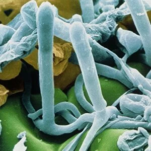
Mushroom underside. Coloured scanning electron micrograph (SEM) of the underside of a chicken of the woods mushroom, also called a sulphur polypore mushroom. This mushroom is the fruiting body of the Laetiporus sulphureus fungus. It is a polypore mushroom that distributes its reproductive spores through the many pores seen here. The pores are up to 3 millimetres long. This polypore mushroom is parasitic on trees, forming large fan-shaped brackets, mostly on oak but also on yew, cherry, willow and sweet chestnut trees. The young, fresh brackets are edible. Magnification unknown
Science Photo Library features Science and Medical images including photos and illustrations
Media ID 6275827
© SUSUMU NISHINAGA/SCIENCE PHOTO LIBRARY
Bracket Edible Eumycota Fruiting Body Fungal Fungi Fungus Mushroom Mycology Naturemycology Parasite Parasitic Pore Pores Re Production Reproductive Surface Under Side Underneath Chicken Of The Woods Laetiporus Sulphureus Sulphur Polypore
MADE IN THE USA
Safe Shipping with 30 Day Money Back Guarantee
FREE PERSONALISATION*
We are proud to offer a range of customisation features including Personalised Captions, Color Filters and Picture Zoom Tools
SECURE PAYMENTS
We happily accept a wide range of payment options so you can pay for the things you need in the way that is most convenient for you
* Options may vary by product and licensing agreement. Zoomed Pictures can be adjusted in the Cart.