Fraud Blocker Skip to main content

Eumycota Collection

Eumycota, the diverse kingdom of fungi, encompasses a fascinating array of organisms

Background imageEumycota Collection: Budding yeast cell

Budding yeast cell. Computer artwork of asection through a yeast ( Candida albicans )cell that is reproducing asexually. A daughtercell (top left) is budding from the parent cell

Background imageEumycota Collection: Fly agaric mushrooms

Fly agaric mushrooms (Amanita muscaria), artwork. This mushroom is powerfully hallucinogenic, containing the drugs ibotenic acid and muscimol

Background imageEumycota Collection: SEM of penicillin fungus

SEM of penicillin fungus

Background imageEumycota Collection: Culture of Aspergillus nidulans fungus

Culture of Aspergillus nidulans fungus
Aspergillus nidulans. Laboratory petri-dish culture of the fungus Aspergillus nidulans, cause of aspergillosis and mycetoma

Background imageEumycota Collection: Cep mushroom (Boletus edulis)

Cep mushroom (Boletus edulis)
Cep mushrooms (Boletus edulis). This fungus is edible

Background imageEumycota Collection: Sporangiophores of Pilobolus fungus on dung

Sporangiophores of Pilobolus fungus on dung
Fungus sporangiophores. Macrophotograph of three sporangiophores of a Pilobolus sp. fungus, growing on dung. A sporangiophore is a fruiting body formed on a specialised hypha

Background imageEumycota Collection: Mushroom gills, SEM

Mushroom gills, SEM
Mushroom gills. Coloured scanning electron micrograph (SEM) of the gills of an unidentified gilled mushroom. The mushroom is the fruiting body of a fungus

Background imageEumycota Collection: Fly agaric fungi

Fly agaric fungi (Amanita muscaria). The mycelium of the fly agaric fungus shares a symbiotic relationship with spruce, forming a mycorrhizal sheath around the roots of the tree

Background imageEumycota Collection: Dividing yeast cells, SEM

Dividing yeast cells, SEM
Dividing yeast cells. Coloured scanning electron micrograph (SEM) of Schizosaccharomyces pombe yeast cells dividing. S. pombe is a single-celled fungus that is studied widely as a model organism for

Background imageEumycota Collection: Dividing yeast cells, SEM

Dividing yeast cells, SEM
Dividing yeast cells. Coloured scanning electron micrograph (SEM) of Schizosaccharomyces pombe yeast cells dividing. S. pombe is a single-celled fungus that is studied widely as a model organism for

Background imageEumycota Collection: Fruiting bodies of Rhizopus oligosporus

Fruiting bodies of Rhizopus oligosporus
Rhizopus oligosporus fungus. Coloured scanning electron micrograph of fruiting bodies and spores of the fungus Rhizopus oligosporus

Background imageEumycota Collection: Pilobolus fungus

Pilobolus fungus
Fungus sporangiophores. Coloured scanning electron micrograph (SEM) of sporangiophores of a Pilobolus sp. fungus, growing on dung

Background imageEumycota Collection: Art of yeast cell budding

Art of yeast cell budding
Illustration of a yeast cell in the process of budding. In this view of the surface of the cell, collar- like outgrowths are seen

Background imageEumycota Collection: LM of Cryptococcus neoformans fungi

LM of Cryptococcus neoformans fungi
Coloured light micrograph of Cryptococcus neoformans fungi, the cause of cryptococcosis. This yeast-like organism grows by budding in infected tissues and in culture at temperatures between 25

Background imageEumycota Collection: Yeast cells, SEM

Yeast cells, SEM
Yeast cells. Coloured scanning electron micrograph (SEM) of cells of brewer s, or baker s, yeast (Saccharomyces cerevisiae). This fungus consists of single vegetative cells

Background imageEumycota Collection: Yeast cells, SEM

Yeast cells, SEM
Yeast cells. Coloured scanning electron micrograph (SEM) of cells of bakers yeast (Saccharomyces cerevisiae) from part of a dried, commercial yeast pellet

Background imageEumycota Collection: Culture of Penicillium chrysogenum fungus

Culture of Penicillium chrysogenum fungus
Penicillium chrysogenum. Laboratory petri- dish culture of the fungus Penicillium chrysogenum growing on agar. Growth is from the centre outwards

Background imageEumycota Collection: Penicillium chrysogenum fungal culture

Penicillium chrysogenum fungal culture

Background imageEumycota Collection: Yeast cells, TEM

Yeast cells, TEM
Yeast cells, coloured transmission electron micrograph (TEM). The cell wall is shown in blue, cytoplasm in red, and vacuole in green. Yeast is a single-celled fungus that reproduces asexually

Background imageEumycota Collection: SEM of yeast cells

SEM of yeast cells
Yeast. Coloured scanning electron micrograph (SEM) of yeast cells, Saccharomyces cerevisiae, commonly known as Bakers or Brewers yeast, growing on potato dextrose agar

Background imageEumycota Collection: Blue stain fungi

Blue stain fungi (Chlorociboria aeruginascens) on rotting wood. This inedible fungus stains the wood it grows on a blue-green colour. Such wood was formerly used for veneers in marquetry

Background imageEumycota Collection: Bread mould, SEM C016 / 9051

Bread mould, SEM C016 / 9051
Bread mould. Coloured scanning electron micrograph (SEM) of a black bread mould (Rhizopus stolonifer) sporangium (orange, spherical)

Background imageEumycota Collection: Bread mould, SEM C016 / 9052

Bread mould, SEM C016 / 9052
Bread mould. Coloured scanning electron micrograph (SEM) of a black bread mould (Rhizopus stolonifer) sporangium (blue, spherical)

Background imageEumycota Collection: Mushrooms spores, SEM C016 / 9450

Mushrooms spores, SEM C016 / 9450
Mushrooms spores. Coloured scanning electron micrograph (SEM) of budding spores in the basidium of an Agaricus bisporus fungus

Background imageEumycota Collection: Bread mould, SEM C016 / 9050

Bread mould, SEM C016 / 9050
Bread mould. Coloured scanning electron micrograph (SEM) of a black bread mould (Rhizopus stolonifer) sporangium (green, spherical)

Background imageEumycota Collection: Penicillium fungal spores, SEM

Penicillium fungal spores, SEM
Penicillium fungal spores. Coloured scanning electron micrograph (SEM) of fruiting bodies of the fungus Penicillium roqueforti

Background imageEumycota Collection: Penicillium spores

Penicillium spores
Penicillium. Coloured scanning electron micrograph (SEM) of the mycelium and fruiting bodies of the fungus Penicillium sp

Background imageEumycota Collection: Fungus on a fly

Fungus on a fly
Fungus on fly. Coloured scanning electron micrograph (SEM) of an entomogenous fungus, Empusa muscae erupting from the abdomen of a housefly (Musca domestica)

Background imageEumycota Collection: Mould sporangia

Mould sporangia. Coloured scanning electron micrograph (SEM) of the sporangia (orange) of an unidentified mould fungus. The sporangia are the organs in which the asexual spores are produced

Background imageEumycota Collection: Hyphae and asci of Sphaerotheca mildew

Hyphae and asci of Sphaerotheca mildew
Mildew. Coloured Scanning Electron Micrograph of hyphae and ripe asci of the leaf mildew Sphaerotheca sp. The hyphae are the tangled filaments which make up a fungal colony

Background imageEumycota Collection: Yeast, Saccharomyces cerevisiae

Yeast, Saccharomyces cerevisiae
Yeast cells. Coloured scanning electron micro- graph (SEM) of budding yeast cells (yellow, Saccharomyces cerevisiae). Known as bakers or brewers yeast

Background imageEumycota Collection: Field Mushroom basidiospores

Field Mushroom basidiospores
Mushroom basidiospores. Coloured scanning electron micrograph (SEM) of basidiospores of the Field Mushroom (Agaricus campestris)

Background imageEumycota Collection: Porcelain mushroom (Oudemansiella mucida)

Porcelain mushroom (Oudemansiella mucida)
Porcelain mushrooms (Oudemansiella mucida) growing on a moss-covered branch. This edible fungus is named after its translucent appearance. Photographed in the New Forest, UK, in October

Background imageEumycota Collection: Magic mushrooms (Psilocybe semilanceata)

Magic mushrooms (Psilocybe semilanceata). These fungi, commonly known as liberty caps, contain the psychoactive compound psilocybin, which produces a hallucinogenic effect when ingested

Background imageEumycota Collection: Many-coloured polypore fungi

Many-coloured polypore fungi
Many-zoned polypore (Coriolus versicolor) bracket fungi. Wiltshire, UK November 2010

Background imageEumycota Collection: Stilton cheese, SEM

Stilton cheese, SEM
Stilton. Coloured scanning electron micrograph (SEM) of a blue vein in a piece of Stilton cheese. The colour and flavour of the cheese is produced by the fungus Penicillium roquefortii

Background imageEumycota Collection: Curvularia geniculata fungus, SEM

Curvularia geniculata fungus, SEM
Curvularia geniculata fungus. Coloured scanning electron micrograph (SEM) of the fruiting body of the mould fungus Curvularia geniculata

Background imageEumycota Collection: Bracket fungus (Laetiporus sulphureus)

Bracket fungus (Laetiporus sulphureus)
Bracket fungus. Young sulphur-coloured fruiting body of the edible bracket fungus known as the chicken of the woods (Laetiporus sulphureus), growing on a living willow tree

Background imageEumycota Collection: Bracket fungus (Daedaleopsis confragosa)

Bracket fungus (Daedaleopsis confragosa)
Blushing bracket fungus (Daedaleopsis confragosa). Close-up of the underside of the blushing bracket fungus showing its pores. The pores are the ends of the tubes within which the spores are produced

Background imageEumycota Collection: Bracket fungus (Polyporus squamosus)

Bracket fungus (Polyporus squamosus)
Bracket fungus. Dryads saddle (Polyporus squamosus) bracket fungus on a tree

Background imageEumycota Collection: Honey fungus

Honey fungus (Armillaria mellea) growing on a tree trunk. Photogroahed in Narke, Sweden

Background imageEumycota Collection: Hares ear fungus

Hares ear fungus (Otidea onotica). Photographed in Narke, Sweden

Background imageEumycota Collection: Birch bolete (Leccinum scabrum) mushroom

Birch bolete (Leccinum scabrum) mushroom
Brown birch bolete (Leccinum scabrum) mushroom growing amongst common heather (Calluna vulgaris). Photographed in the Cairngorms National Park, Scotland, in August

Background imageEumycota Collection: Fly agaric (Amanita muscaria) mushrooms

Fly agaric (Amanita muscaria) mushrooms growing amongst common heather (Calluna vulgaris) in an ancient Caledonian Scots Pine woodland

Background imageEumycota Collection: Penicillium fungus, SEM

Penicillium fungus, SEM
Penicillium fungus. Coloured scanning electron micrograph (SEM) of a Penicillium sp. fungus. Specialised threads (hyphae, pink), called conidiophores, are seen

Background imageEumycota Collection: Amanita (Amanita frostiana) mushroom

Amanita (Amanita frostiana) mushroom
Frosts amanita (Amanita frostiana) mushroom. Photographed in Vermont, USA

Background imageEumycota Collection: Woolly gomphus (Gomphus floccosus)

Woolly gomphus (Gomphus floccosus) mushroom. Photographed in Vermont, USA

Background imageEumycota Collection: Dry rot fungus

Dry rot fungus. Dry rot, unlike its name suggests, infects damp timber. The term refers to the growth of either of two fungi


All Professionally Made to Order for Quick Shipping

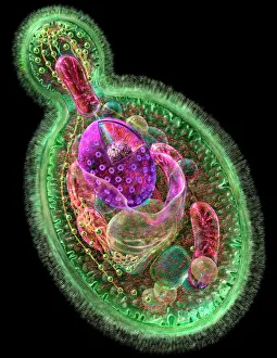
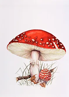
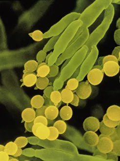
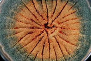
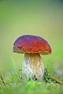
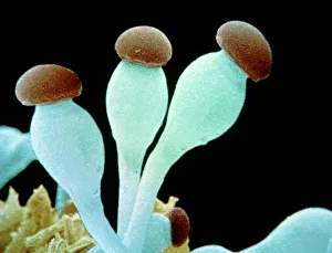
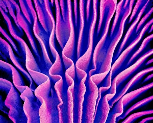
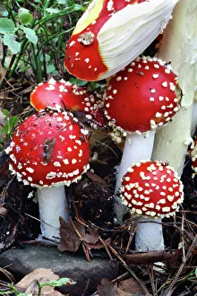
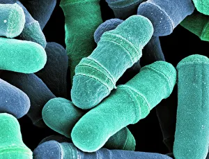
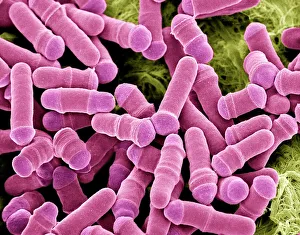
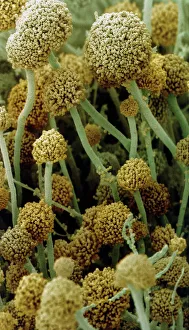
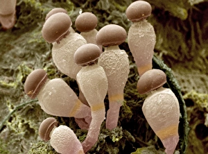
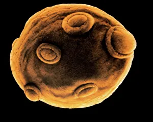
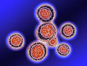
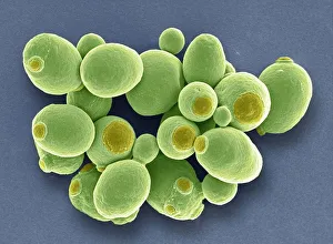
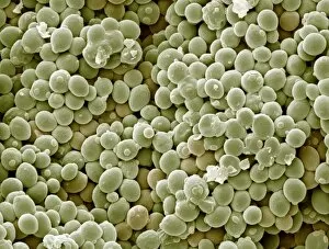
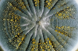
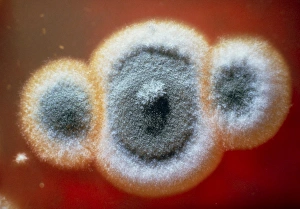
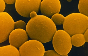
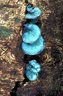
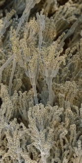
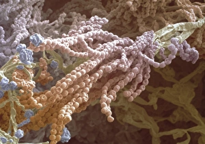
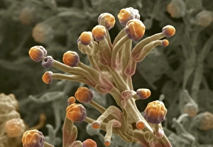
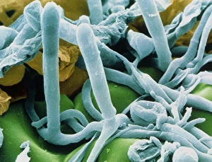
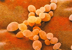
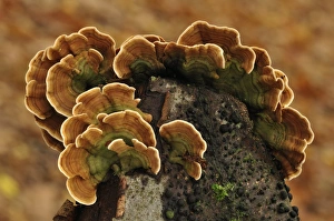
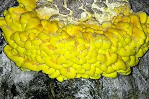
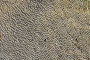
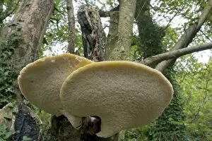
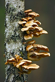
Eumycota, the diverse kingdom of fungi, encompasses a fascinating array of organisms. From the microscopic world of budding yeast cells and dividing yeast cells seen through scanning electron microscopy (SEM), to the enchanting fly agaric mushrooms with their distinctive red caps and white spots, this caption explores some captivating members of Eumycota. In our first glimpse into this kingdom, we encounter the SEM image of penicillin fungus - a remarkable organism that has revolutionized medicine by producing antibiotics. Moving on to Aspergillus nidulans fungus in culture, its intricate structures captivate us as it thrives in laboratory conditions. Nature's artistry is showcased through the Cep mushroom or Boletus edulis - an edible delicacy prized for its rich flavor and meaty texture. The SEM image of mushroom gills reveals their intricate network responsible for spore production and dispersal. Fly agaric fungi transport us into fairy tales with their vibrant colors and whimsical appearance. These iconic mushrooms have long been associated with folklore and mythical creatures. Returning to the microscopic realm, we witness dividing yeast cells under SEM once again - a testament to their incredible ability to reproduce rapidly. Penicillium roqueforti fungus takes center stage next; known for its role in aging blue cheeses like Roquefort, it adds distinct flavors as it grows within these culinary delights. Fruiting bodies of Rhizopus oligosporus catch our attention next – these structures are essential for reproduction in this species while Cryptococcus neoformans fungi reveal themselves under light microscopy (LM). This pathogenic fungus can cause severe infections particularly in individuals with compromised immune systems. Eumycota truly showcases nature's diversity at both macroscopic and microscopic levels. Whether they provide sustenance or pose health risks, these organisms remind us how intricately interconnected our world is with fungi playing vital roles across various ecosystems.