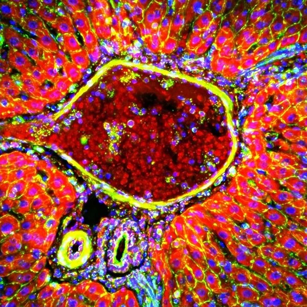
Liver portal triad, light micrograph C016 / 8489
![]()

Wall Art and Photo Gifts from Science Photo Library
Liver portal triad, light micrograph C016 / 8489
Liver portal triad. Fluorescence deconvolution micrograph of a section through a portal triad in liver tissue, showing hepatocyte cells (red), connective tissue (green) inside the periportal limiting plate, and cell nuclei (blue dots). A portal triad (portal field, or portal tract) is a distinctive arrangement in the liver where the proper hepatic artery, hepatic portal vein, common bile duct, lymphatic vessels and a branch of the vagus nerve meet. The ring of hepatocytes abutting the connective tissue of the triad is called the periportal limiting plate
Science Photo Library features Science and Medical images including photos and illustrations
Media ID 9221949
© R. BICK, B. POINDEXTER, UT MEDICAL SCHOOL/SCIENCE PHOTO LIBRARY
Animal Tissue Bile Duct Cell Biology Confocal Light Micrograph Connective Tissue Cytological Cytology Dyes Fluorescence Fluorescent Deconvolution Hepatic Hepatic Portal Vein Hepatocyte Hepatocytes Histological Histology Liver Lymph Marker Markers Nuclei Nucleus Proteins Rat Tissue Rodent Stain Stains Vagus Nerve Vessels Blood Vessel Cells Light Micrograph Light Microscope Protein Section Sectioned
EDITORS COMMENTS
This print showcases the intricate details of a liver portal triad, providing a glimpse into the complex biology and anatomy of this vital organ. The image, captured using fluorescence deconvolution microscopy, reveals a section through the portal triad in liver tissue. Highlighted in vibrant red are the hepatocyte cells, which play a crucial role in metabolic processes within the liver. Surrounding these cells is green connective tissue found inside the periportal limiting plate, emphasizing its structural significance. Blue dots represent cell nuclei scattered throughout the image, symbolizing their presence and importance within this microcosm. A portal triad is an extraordinary arrangement where various components converge: the proper hepatic artery, hepatic portal vein, common bile duct, lymphatic vessels, and a branch of the vagus nerve. This convergence forms a ring of hepatocytes known as the periportal limiting plate that abuts against connective tissue. The photograph offers valuable insights into normal liver anatomy and function while showcasing its biological complexity at a microscopic level. It serves as an invaluable resource for researchers studying liver histology or cytology by providing detailed visual information about cellular structures and protein markers present in healthy animal tissues such as rat livers. Captured by R. BICK and B. POINDEXTER from UT Medical School's Science Photo Library collection using advanced light microscopy techniques like fluorescent deconvolution imaging; this stunning print represents both scientific excellence and aesthetic beauty simultaneously
MADE IN THE USA
Safe Shipping with 30 Day Money Back Guarantee
FREE PERSONALISATION*
We are proud to offer a range of customisation features including Personalised Captions, Color Filters and Picture Zoom Tools
SECURE PAYMENTS
We happily accept a wide range of payment options so you can pay for the things you need in the way that is most convenient for you
* Options may vary by product and licensing agreement. Zoomed Pictures can be adjusted in the Cart.

