
Bile Duct Collection
The bile duct, a vital component of our digestive system, plays a crucial role in the transportation of bile from the liver to the small intestine


All Professionally Made to Order for Quick Shipping
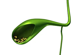
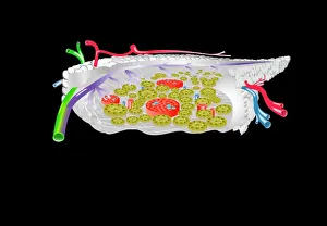
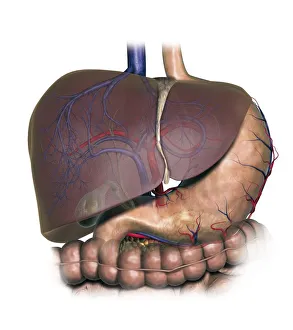
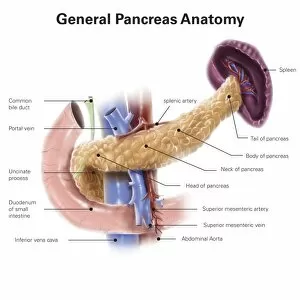
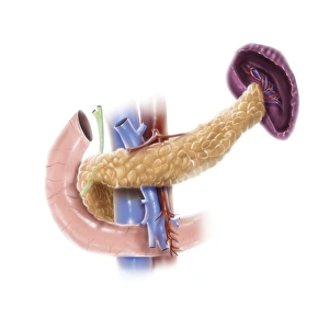
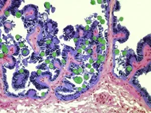
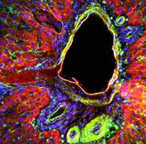
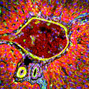
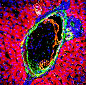
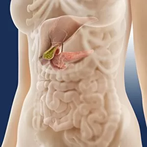
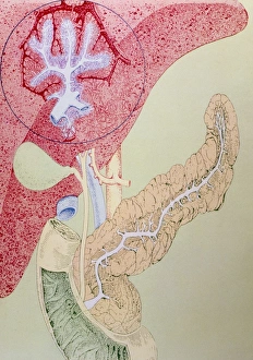
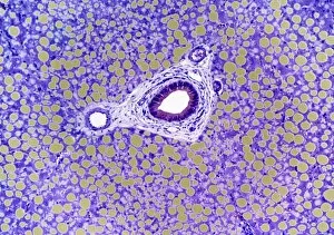
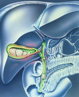
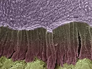
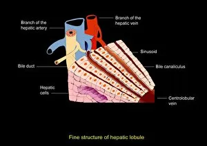
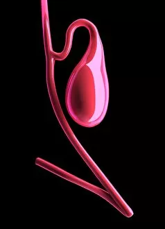
The bile duct, a vital component of our digestive system, plays a crucial role in the transportation of bile from the liver to the small intestine. This intricate network is often affected by gallstones, causing discomfort and requiring medical intervention such as cholecystectomy, the surgical removal of the gallbladder. Intriguing artwork showcases the complexity of pancreas anatomy, shedding light on its connection to both digestion and hormone regulation. A cross-section biomedical illustration vividly depicts how this organ interacts with other components of our digestive system like the esophagus. A detailed diagram reveals an interconnected web within our bodies, showcasing not only the liver but also highlighting key players like stomach and gallbladder alongside pancreas. With labeled sections providing insight into their functions, it becomes evident how each element contributes to overall well-being. Conceptual images capture gallstones inside a gallbladder - tiny yet capable of causing significant distress if left untreated. These visual representations emphasize why understanding bile duct health is essential for maintaining optimal digestive function. Additionally, microscopic views offer glimpses into coccidiosis - an infection that can affect various organs including bile ducts. Light micrographs F005 / 6082 and F005 / 6081 provide fascinating insights into this condition's impact on these delicate structures. From informative illustrations to captivating artwork, exploring different aspects related to bile ducts allows us to appreciate their significance in maintaining a healthy digestive system. Whether it's understanding their anatomy or recognizing potential issues like gallstones or infections such as coccidiosis – delving deeper into this subject unveils just how remarkable our bodies truly are.