Home > Arts > Artists > B > C.L. Boes
Heart chamber wall defect, artwork

![]()
Wall Art and Photo Gifts from Science Photo Library
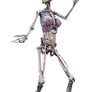
Heart chamber wall defect, artwork
Heart anatomy. Artwork of a posterior (from behind) view of a normal heart, sectioned to show the internal anatomy. The red arrow shows the normal flow of blood from the left ventricle (at left) into the ascending aorta (curving to upper left). at right is the right ventricle. Above the ventricles are the other two heart chambers, the left and right atria respectively. Between each atrium and ventricle are atrioventricular valves (white): the mitral valve (left) and the tricuspid valve (right). The left ventricle has particularly thick walls of cardiac muscle (myocardium), because it is this chamber that pumps blood around the body
Science Photo Library features Science and Medical images including photos and illustrations
Media ID 9224445
© BO VEISLAND/SCIENCE PHOTO LIBRARY
Cardiac Cardiology Cardiovascular Chordae Tendineae Left Ventricle Mitral Valve Right Ventricle Tricuspid Valve Condition Cutouts Disorder Health Care Semilunar Valve
FEATURES IN THESE COLLECTIONS
> Arts
> Artists
> B
> C.L. Boes
EDITORS COMMENTS
This artwork captures the intricate internal anatomy of a normal heart, showcasing its various chambers and valves. From a posterior view, we can observe the left ventricle on the left side of the image, distinguished by its thick walls of cardiac muscle. This chamber plays a crucial role in pumping oxygenated blood throughout the body. The red arrow indicates the normal flow of blood from this ventricle into the ascending aorta, which curves towards the upper left. On the right side of the image lies the right ventricle, responsible for pumping deoxygenated blood to be reoxygenated in the lungs. Above both ventricles are two additional heart chambers known as atria – specifically, on top is the left atrium and below it is the right atrium. These chambers are separated from their corresponding ventricles by atrioventricular valves: namely, on our left is mitral valve and on our right is tricuspid valve. The detailed illustration also highlights other significant features such as chordae tendineae (tendinous cords), semilunar valves (notably absent in this particular view), papillary muscles, and even depicts potential congenital defects like ventricular septal defect (VSD) associated with conditions like Tetralogy of Fallot or blue baby syndrome. Overall, this stunning print provides an insightful glimpse into one's cardiovascular system while emphasizing both healthy anatomy and potential abnormalities that may affect heart function.
MADE IN THE USA
Safe Shipping with 30 Day Money Back Guarantee
FREE PERSONALISATION*
We are proud to offer a range of customisation features including Personalised Captions, Color Filters and Picture Zoom Tools
SECURE PAYMENTS
We happily accept a wide range of payment options so you can pay for the things you need in the way that is most convenient for you
* Options may vary by product and licensing agreement. Zoomed Pictures can be adjusted in the Cart.